Meniscal tears and meniscus rupture
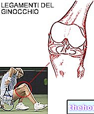
The most common knee injuries are those affecting the menisci, two small C-shaped fibrocartilaginous structures located between the femoral condyles and the tibia. The menisci allow a better distribution of the loads on the articular cartilage, attenuating them and at the same time guaranteeing the correct mechanics of movement.

During a trivial movement or following a trauma the meniscus can get pinched between the tibia and the femur, tearing itself as a piece of cloth stuck in a door would do.
Fortunately, our body is much more efficient and resistant than any mechanical gear designed by man even if, unfortunately, the regenerative capacity of the menisci is very low. These structures, in fact, despite being quite vascularized at the extremities, have a large central portion devoid of capillaries. Without blood the cells of the injured menisci cannot heal and heal. If we exclude the cases in which the lesion is limited and extended only to one "extremity, a ruptured meniscus therefore has no regenerative capacity.
Classification and causes of meniscal tears
Meniscal tears can be classified into two large groups:
Meniscal lesions of traumatic origin: they are more frequent among young people and sportsmen. In these cases, one or both menisci undergo injuries following a violent stress that supersedes the maximum resistance of the cartilage tissue that composes them.
Meniscal lesions of degenerative origin: the meniscus is injured following a seemingly trivial movement such as getting up quickly from a squatting position. These lesions arise due to the degeneration of the meniscal tissue which, over the years, becomes more fragile and less elastic.
The lesion can practically affect any point of the meniscus. Ruptures limited to the anterior horn alone are however quite rare. Usually the lesions initially affect the posterior horn and then eventually extend to the central body and the anterior horn. Ligament tears are often associated with these injuries, particularly when the medial or internal meniscus is involved. The injury of this meniscus is about five times more frequent than that of the lateral meniscus due to its greater degree of mobility.
CAUSES: The meniscus is particularly vulnerable when compressive forces associated with twisting forces are applied to it. It follows that most traumatic events occur when the knee undergoes a torsional trauma. If the trauma is applied when the joint is externally rotated (external rotation) there is a greater risk of injuring the medial meniscus and vice versa.
At other times, a meniscal tear occurs as a result of hyperflexion or hyperextension movements, for example by giving a hollow kick.
As we have seen, the meniscal fibrocartilages lose part of their elasticity over time and are more subject to wear. For this reason, many meniscal tears in the elderly are the result of insignificant trauma, such as the act of squatting. A bit like old shirts worn by frequent washing, even the menisci can be torn during habitual movements.
Symptoms
The main symptoms of meniscal tears include pain and local swelling. These two symptoms are often associated with the collapse and blockage of the joint caused by the meniscus fragments that interfere with the normal mobility of the knee.
The pain increases in the position that generated the meniscal tear, for example during its rotation or pressure. Following a meniscal injury, the subject complains:
- inability to fully extend or flex the joint
- inflammation of the membrane leads to increased production of fluid that collects in the joint cavity (hydrarct)
- joint crunch associated with pain
SYMPTOMS for clinical diagnosis:
- pain evoked during particular movements: in the event of a medial meniscus injury, the pain is mainly localized in the internal part of the knee during hyperflexion, hyperextension or external rotation with the knee flexed to 90 °; for the lateral meniscus the opposite is true ( pain localized externally in hyperextension, hyperflexion or internal rotation of the leg and foot with the knee flexed between 70 ° and 90 °)
- loss of strength or hypotrophy of the quadriceps
Diagnosis
The diagnosis of a meniscal tear is fundamentally clinical. The doctor, in his surgery, will look for the presence of the diagnostic symptoms described above. If at least three signs are present at the same time, the diagnosis of meniscus injury, lateral or medial depending on the case, is considered almost certain.
In any case, the diagnosis must be confirmed by an instrumental investigation.
The x-ray does not provide direct information on the health of the meniscus, since this is not a calcified structure, but it can still be useful to exclude other pathologies (osteoarthritis).
Magnetic resonance imaging, on the other hand, is able to provide clear information on the state of soft tissues, including the menisci. Thanks to these characteristics, MRI can highlight any degenerative processes before the meniscus breaks.
CT also provides useful information but less precise and detailed than MRI. This technique is less expensive, has shorter waiting lists, shows bone health very well but provides little information about the menisci.
Finally, we recall arthroscopy, which despite being invasive, represents the safest method to confirm the diagnosis of meniscal injury.
CONTINUE: Treatment of meniscal tears "


