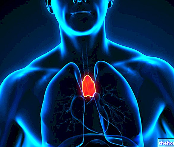
The trachea is an elastic and flexible structure, comparable to a flattened cylinder in the posterior face. From a physiological point of view, it has the purpose of conveying the air from the outside towards the lungs during inspiration and in the opposite direction during exhalation.

About 12 cm long for an average diameter of 2 cm, the trachea connects the larynx to the bronchi. Above it originates from the cricoid cartilage of the larynx, while in the lower part it ends with a bifurcation from which the two primary bronchi arise. From this level onwards, the respiratory tree continues with a dense network of branches: from the primary bronchi originate the secondary bronchi (lobar bronchi) and from these the tertiary bronchi (segmental bronchi), which in turn divide into bronchioles, then in terminal bronchioles and finally in respiratory bronchioles rich in alveoli.
The trachea is formed by a series of overlapping cartilaginous rings, similar to a horseshoe, open in the posterior region and connected to each other by connective tissue.

At the back, the trachea relates to the esophagus, while laterally it relates to the nervous vascular bundle of the neck. From the didactic point of view, it can be divided into two parts. The first, the Pars cervicalis (extrathoracic) continues above with the cartilage cricoid of the larynx (located in the lower part of this organ), extending from the 4th to the 7th cervical vertebrae. Inferiorly, the pars cervicalis continues with the intrathoracic tracheal segment (Pars thoracic), which in turn ends at the limit of the body and manubrium of the sternum (at the level of the IV-V thoracic vertebrae in the adult) dividing into the two primary bronchi.
Due to the particular arrangement of the tracheal rings, from the morphological point of view the trachea appears flattened at the back and rounded in its anterior part.
The anterior-posterior diameter is about 1.5 cm, while the transverse one is around 1.8 cm.
Like all cartilage structures, each tracheal ring is lined with a layer of connective tissue rich in blood vessels and nerve endings, called the perichondrium. The nutritional exchanges of cartilage cells depend on it.

The wall of the trachea, proceeding from the outside towards the inside, has three layers: the adventitious tunic, the submucosa and the mucosa. Without going into anatomical details, let us briefly recall that the mucous membrane of the trachea (see image on the side) is covered by a pseudostratified ciliated cylindrical epithelium (respiratory epithelium), on which a layer of mucus is deposited.
Thanks to the ciliary movements and the adhesive action of the mucus, the trachea is able to "self-clean", trapping foreign agents (dust, pollen, bacteria, etc.) and favoring their elimination. In fact, the tracheal cilia, moving from the bottom upwards, make the mucus rise up to the oral cavity, then towards the esophagus and from there to the stomach, where it is digested by the gastric juices.

