Thanks to these cylindrical units, the chemical energy released by metabolic reactions is transformed into mechanical energy; by inserting itself through the tendons and acting on the bone levers, the muscle generates movement.
Skeletal muscle fibers vary in length from a few millimeters to several centimeters, with a diameter ranging from 10 to 100 µm (1 µm = 0.001 mm); they are the largest cells in the body.
"Cytologically" speaking, fiber cells are the result of a process called myogenesis, which is the fusion of multiple myoblasts - an action dependent on muscle-specific proteins known as fusogens, myomaker or myomerger. This is why myocells appear as long cylindrical and polynucleted cells (which contain numerous myonuclei - among other things, clearly visible on the surface under the microscope).
A muscle fiber, eg. in the brachial biceps, with a length of 10 cm it can have up to 3000 nuclei.
Inside them there are instead thousands of filaments, called myofibrils, containing contractile units called sarcomeres.
Physiologists who deal with muscles tell us that the various fibers differ from each other, not only from the anatomical point of view, but also for some precise physiological characteristics.
Therefore, within each muscle different types of fibers are recognized, classified according to different criteria such as energy metabolism, speed of contraction, resistance to fatigue, color, etc.
Overall, a single muscle such as eg. the brachial biceps, about 253,000 muscle fibers are contained.
Did you know that ...
Between the basement membrane and the sarcolemma of muscle fibers lies a group of muscle stem cells known as myosatellite cells.
These are normally quiescent but can be activated by exercise or disease to provide additional myonuclei needed for muscle growth or repair.
specific, phosphages (ATP and CP), mitochondria, myoglobin, glycogen and a higher capillary density.
However, muscle cells cannot divide to produce new cells and, as a result, their number tends to decrease with age.
), which give rise to three types of fibers.
These fibers have relatively distinct metabolic, contractile and motor properties - summarized in the table below.
IMPORTANT! The various properties, although they depend in part on the characteristics of the individual fibers, tend to be more relevant when measured at the level of the motor unit - which, however, show very minimal variations in terms of variety of fibers - rather than of the single fiber.
Let's now see some types of classification.
Fiber color
Traditionally, fibers were classified according to their color, which depends on the myoglobin content.
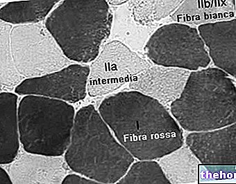
Type I fibers appear red due to high myoglobin levels, tend to have more mitochondria and higher local capillary density.
They are slower to shrink but more suited to resistance, because they use oxidative metabolism to generate ATP (adenosine triphosphate) from glucose and fatty acids.
The less oxidative type II fibers are white or in any case clear, due to the scarcity of myoglobin and the concentration of glycolytic enzymes.
Speed of contraction
Fibers can be classified according to their fast and slow contractile speeds. These traits overlap largely, but not completely, with classifications based on color, ATPase, and MHC.
- Fibers a rapid contraction those in which myosin can split ATP very quickly. These include type II ATPase and type II MHC fibers. They also demonstrate a greater capacity for electrochemical transmission of action potentials and a rapid level of calcium release and absorption by the sarcoplasmic reticulum. They are based on a well developed, anaerobic, fast energy transfer glycolytic system, and can contract 2- 3 times faster than slow twitch fibers Fast twitch muscles are suitable for generating short bursts of strength or speed than slow muscles, and therefore fatigue faster.
- Fibers a slow contraction generate energy for the resynthesis of ATP through an aerobic and long-lasting transfer system. These mainly include ATPase type I and MHC type I fibers. They tend to have a low level of ATPase activity, a slower twitch rate with a less developed glycolytic capacity. Slow twitch fibers develop more mitochondria and capillaries, which makes them better for endurance work.
Fiber typing methods
There are a number of methods employed for fiber typing, which often creates some confusion among non-experts.
Two often equivocal methods are histochemical staining for myosin ATPase activity and immunohistochemical staining for the myosin heavy chain type (MHC).
The activity of the myosin ATPase enzyme is commonly and correctly referred to simply as the "fiber type" and results from the direct measurement of the activity of the ATPase enzyme under various conditions (eg pH).
The myosin heavy chain staining is more precisely referred to as "MHC type" (myosin heavy chain) and, as can be understood, results from the determination of different MHC isoforms.
These methods are physiologically related, since the MHC type is the main determinant of ATPase activity. However, none of these typing methods are directly metabolic in nature; that is they do not directly address the oxidative or glycolytic capacity of the fiber.
When referring to "type I" or "type II" fibers, this refers more accurately to the staining assessment of the "ATPase activity of myosin (e.g." type II "fibers refer to type IIA + type IIAX + type IIXA ... etc.).
Below is a table showing the relationship between these two methods, limited to the types of fibers present in humans.Subtype capitalization is used in fiber typing versus MHC typing; some types of ATPase actually contain multiple types of MHC.
Furthermore, a subtype B or b is not expressed in humans by either method. Early researchers believed that humans could express an MHC IIb, which led to the ATPase classification of IIB. However, subsequent research has shown that human MHC IIb is actually IIx, indicating that the more correct wording is IIx.
Subtype IIb or IIB, IIc and IId, are instead expressed in other mammals, as is widely documented in the literature.
Further fiber typing methods are outlined in a less formal way and exist on more spectra, such as the one normally used in the athletic-sports field.
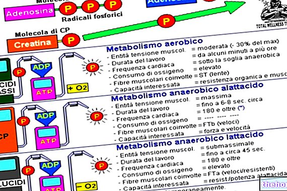
They tend to focus more on metabolic and functional capacities (contraction time, predominantly oxidative vs. anaerobic lactacid vs. anaerobic lactacid, fast vs. slow contraction time).
As noted above, fiber typing by ATPase or MHC does not directly measure or dictate these parameters. However, many of the various methods are mechanically linked, while others are related in vivo.
Eg, the type of ATPase fiber is related to the speed of contraction, since the high activity of ATPase allows a faster cycle of the cross bridge. Type I fibers are "slow", in part, because they have low rates of ATPase activity compared to type II fibers; however, measuring the rate of contraction is not the same as typing the ATPase fiber.
, white and intermediate fibers. Their proportions, however, vary according to the physiologically assigned work to that muscle.For example, in humans, the quadriceps muscles contain about 52% of type I fibers, while the soleus is about 80%. The orbicularis muscle of the eye, on the other hand, has only about 15% of type I.
Did you know that ...
The force developed by a muscle fiber depends on its length at the beginning of the contraction. It must have an optimal value, outside of which (retracted or excessively stretched muscle) strength performance is reduced. In the field of muscle strengthening, the most common mistake is to work the muscles already in partial shortening. The only exceptions to the rule are the presence of pain or discomfort, or paramorphisms, which therefore require a limitation of the range of motion (ROM).
The predominantly white muscles, rich in type II fibers, are called phasic, because they are capable of rapid and short contractions. The red muscles, on the other hand, where type I fibers prevail, are called tonic, due to the ability to remain in contraction for a long time.
The motor units within the muscle, however, show very little variation, making the dimensional principle of motor unit recruitment; that is, depending on the intensity / strength required, the body is able to stimulate only some (eg in prolonged aerobic activity) or all (eg during a maximal squat) the units in question.
Today we know that there are no sex-related differences in the distribution of fibers. However, the proportions of the various types - which we know vary greatly between animal species and to a lesser extent between ethnicities - "could" vary considerably from person to person.
According to some insights, sedentary men and women (as well as young children) should have 55% type I fiber and 45% type II fiber.
High-level athletes, on the other hand, have a specific fiber distribution based on the type of metabolism used. Cross-country skiers have mainly fibers I, sprinters mainly II and middle-distance runners, throwers and jumpers, almost overlapping percentages of both.
It has therefore been suggested that various types of exercise can induce significant changes in skeletal muscle fibers, although it is not possible to establish with certainty what the pre-existing genetic makeup of the same subjects was. This process "could" be allowed by the specialization capacity of the fibers, or even just a part, belonging to macro-set II.
It is possible that type IIx fibers show improvements in oxidative capacity following high intensity endurance training, leading them to a level where they would become capable of fulfilling oxidative metabolism as effectively as fibers I in untrained subjects.
This would be determined by an increase in the size and number of mitochondria and their associated changes, but not by a change in the type of fiber..



.jpg)


