Key points
Necrotizing fasciitis is a severe, violent and sudden infection of the soft tissues, with a predominantly bacterial etiology.
Necrotizing fasciitis: causes
The bacteria most involved in necrotizing fasciitis are: group A β haemolytic streptococcus, staphylococci (especially Staphylococcus aureus), anaerobes belonging to the genus Clostridium, Vibrio parahaemolyticus, Vibrio vulnificus, Aeromonas hydrophila.
Necrotizing fasciitis: symptoms
The most recurrent signs and symptoms in necrotizing fasciitis include: skin redness, chills, weakness, diarrhea, limited pain, edema, fever, bruising, tissue necrosis, shock, sweating, vomiting. If left untreated, tissue necrosis gives a poor prognosis.
Necrotizing fasciitis: therapy
Therapy for necrotizing fasciitis must be immediate and consists in the administration of antibiotics in high doses and in the surgical excision of the infected tissues. Intensive supportive therapy and the hyperbaric chamber are also useful.
Definition
Fortunately rare, necrotizing fasciitis is a severe soft tissue infection, usually caused by toxigenic bacteria. Necrotizing fasciitis affects the deeper layers of the skin, rapidly spreading through the superficial and deep fascia of the subcutaneous soft tissues.
- Any deep soft tissue compartment - dermis, subcutaneous tissue, muscle bundles - is a possible target for this potentially deadly infection. However, necrotizing fasciitis has a marked preference for the lower limbs, perineum and abdominal wall.
Necrotizing fasciitis is a sudden onset disease that must be cured within the shortest time possible with high doses of intravenous antibiotics.
Necrotizing fasciitis is also known by many other names, which give an immediate indication of the severity of the infection: cellulite acute gangrenous, meat-eating disease, meat-eating bacteria syndrome. Depending on the location of the infection, necrotizing fasciitis takes different names: Fournier's gangrene (necrotizing fasciitis of the scrotum and vulva) e Ludwig's angina (necrotizing fasciitis of the submandibular space).
Necrotizing fasciitis is a very rare infection but has a very high mortality rate.
Causes and classification
Necrotizing fasciitis is caused by bacterial (predominantly) and fungal (rare) infections.
From the etiopathological point of view, several different entities are recognized:
- TYPE I NECROTIZING FASCITIS: polymicrobial infection caused also and above all by type A streptococci (Streptococcus pyogenes), C and G. This form of necrotizing fasciitis particularly affects immunocompromised or chronic disease patients.
- TYPE II NECROTIZING FASCITIS: monomicrobial infection, carried in particular by group A streptococcus, staphylococci or anaerobes belonging to the genus Clostridium (eg. Clostridium perfringens). Also Methicillin-resistant Staphylococcus aureus (MRSA) is involved in this variant of necrotizing fasciitis.
- TYPE III NECROTIZING FASCITIS: serious infections carried by marine microorganisms, such as Vibrio parahaemolyticus, Vibrio vulnificus and Aeromonas hydrophila. Subjects suffering from liver disease are the most sensitive to this form of necrotizing fasciitis: these infections are particularly virulent and fatal (if not treated promptly, death occurs within 48 hours of the onset of the first symptoms).
- TYPE IV NECROTIZING FASCITIS: fungal infections. Patients with traumatic wounds or burns are more at risk of getting infections from Zygomycetes; the immunocompromised are more exposed to mycoses caused by Candida albicans.
From medical statistics, it is clear that necrotizing fasciitis occurs more often in some categories of patients: diabetics, drug addicts, alcoholics, vascular and immunocompromised patients in general. Among other risk factors, we also remember: tuberculosis, malignant neoplasms, infections with Herpes zoster (the virus responsible for chickenpox and St. Anthony's fire).
However, healthy subjects are not exempt from the disease.
Symptoms
Symptoms of necrotizing fasciitis usually manifest within a few days. The symptomatological picture precipitates in 48 hours in patients infected with Vibrio spp. And Aeromonas hydrophila: in such situations, death looms in a few hours.
In general, the symptoms of necrotizing fasciitis vary over time: the more the disease evolves, the more the symptoms get worse. Let's see in detail the course of the disease:
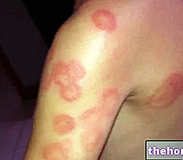
- In the first two days after the infection, the patient complains of limited and constant PAIN, ERITEMA and SWELLING. This symptomatic triad is easily confused with the characteristic signs of erysipelas and infectious cellulitis. The margins of the infection are not well defined and the particular "SOFTNESS" "OF THE SKIN extends beyond the point of infection. During this phase, the disease does not respond to antibiotic treatments. LYMPHANGITIS (inflammation of the lymphatic vessels) is rarely observed. Other symptoms include: TACHY, FEVER, DEHYDRATION , DIARREA and VOMIT.
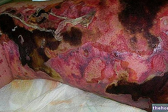
- After 2-4 days, necrotizing fasciitis causes EDEMA, DIFFUSED ERITEMA, BUBBLE LESIONS and hemorrhagic. The skin, first reddened, takes on a greyish color, synonymous with necrosis. The skin tissues are rigid and tense to the touch, while the muscle bundles are no longer palpable. At this stage, many patients no longer feel pain, as necrotizing fasciitis is destroying the nerves.
- On the fourth / fifth day, the patient experiences HYPOTENSION, CONFUSION, APATHY and SEPTIC SHOCK.
If not promptly treated, necrotizing fasciitis is FATAL for 73% of patients.
Diagnosis
The diagnosis of necrotizing fasciitis consists in "medical observation of the lesions. In the case of suspected necrotizing fasciitis, the patient undergoes CT, blood analysis and biopsy of a portion of injured tissue. In addition to the diagnostic techniques listed, we often resort to a immediate exploratory surgery, useful for both diagnostic and therapeutic purposes: after having ascertained necrotizing fasciitis, a large infected tissue portion is removed immediately. In the event that the infection spreads to peripheral regions, the limb is amputated.
Differential diagnosis
Necrotizing fasciitis is quite complex to diagnose in its early stages: this infectious form is often mistaken for bacterial cellulitis. Diagnostic delay postpones therapy, consequently the risk of a fatal outcome increases exaggeratedly. For the differential diagnosis it is important to focus attention on some parameters which, in infectious cellulite, are not very evident or even absent:
- Obvious softness of the affected skin
- Excessive pain, which is accentuated to the touch
- Blisters and bruising on the skin near the site of infection
Therapy
Treatment for necrotizing fasciitis includes:
- Surgical therapy: it consists in the removal of infected tissue flaps, up to the limb amputation. Given the delicacy and complexity of the operation, the patient is generally subjected to multiple surgeries, possibly associated with skin and tissue transplantation.
- Administration of high-dose antibiotics: Antibiotic therapy is also indicated in case of suspected necrotizing fasciitis. The therapy consists of a mix of antibiotics, of which penicillin, clindamycin and vancomycin appear to be the most effective.
- Intensive supportive therapy: useful for dealing with hypotension, the violent inflammatory response of the organism and septic shock. Here, the patient with necrotizing fasciitis undergoes a transfusion of fluids and blood.
- Hyperbaric oxygen therapy: therapy strategy indicated for all patients suffering from tissue destruction and extensive wounds.
Immediate intervention is necessary to prevent the fatal outcome of the patient with necrotizing fasciitis.

-cause-sintomi-e-cura.jpg)
