With still unknown causes, vertebral angioma is only rarely a symptomatic condition; specifically, in such circumstances, the typical symptoms are: back pain, muscle weakness, numbness and numbness in the lower limbs, paralysis of different body areas, loss of control of the anal and bladder sphincters and deformation of the spinal column.
An accurate diagnosis of vertebral angioma involves doing an X-ray and MRI of the spine, as well as an angiography.
Planning and conducting treatment for vertebral angioma only takes place when the condition is symptomatic.
Brief review of what "is a hemangioma
A hemangioma is a particular benign tumor, belonging to the category of angiomas, which originates from the abnormal proliferation of a typical endothelial cell of the inner wall of blood vessels.
Made up of a dense agglomeration of capillaries and slightly larger blood vessels, a hemangioma can:
- Appear as a smooth patch, raised papule, or thick lump
- Be red or purple in color;
- Reside on the skin (cutaneous localization), in the dermis, on the mucous membranes or internal organs (brain, heart, spleen, liver, respiratory tract, bones, etc.).
According to the most recent classifications, there are three types of hemangioma: capillary hemangioma, cavernous hemangioma and pyogenic granuloma (for their characteristics, see the table below).
- Skin
- Smooth patch or raised papule
- Red or purple color
- Dermis
- Internal organs
- Nodule
- Red or purple color
- Skin
- Mucous membranes
- Nodule
- Red
where the benign tumor is located. It is the result of the compression that the vertebral angioma exerts on the spinal cord, contained within the vertebra on which the benign tumor in question is located.
If the vertebral angioma is particularly large or grows larger over time, the painful sensation may spread elsewhere and affect the upper (arms and hands in particular) and lower limbs (especially hips, legs and feet).
Factors that make a vertebral angioma a symptomatic presence:
- Large size of the tumor mass
- Compression of the spinal cord
- Compression of the sensory spinal nerves
- Infiltration of the benign tumor on the vertebra that hosts it and consequent vertebral collapse
When to see a doctor?
In general, the sudden onset of symptoms, such as back pain, a sense of muscle weakness and numbness in the lower limbs, loss of control of the anal sphincters, is always a valid reason to contact your doctor immediately and schedule a check-up. and bladder, loss of sensation in the lower limbs and paralysis of some parts of the body.
For people with asymptomatic vertebral angioma, the recommendation is to consult a doctor experienced in spinal pathologies when the benign tumor begins to manifest itself with the aforementioned symptoms.
Complications
The complications of a vertebral angioma obviously concern only the symptomatic cases and consist in a worsening of the symptoms, such as to seriously compromise the quality of life of the patients.
Regarding a possible malignant evolution of the vertebral angioma (ie the transformation of the vertebral angioma into a malignant tumor of the spine), this feared phenomenon is very rare, if not impossible.
How to diagnose symptomatic vertebral angioma
Canonically, the diagnosis of symptomatic vertebral angioma is based on: a thorough physical examination, a thorough medical history, x-ray of the spine, nuclear magnetic resonance of the spine and angiography (or, alternatively, angioTAC).
OBJECTIVE EXAMINATION AND HISTORY
Physical examination and anamnesis essentially consist in the observation and critical study of the symptoms manifested by the patient.
They always constitute the first step in the process of examinations and tests that lead to the diagnosis of vertebral angioma.
RADIOGRAPHY AND NUCLEAR MAGNETIC RESONANCE OF THE SPINE
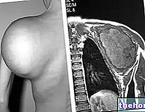
Although with different technologies and physical principles, radiography and nuclear magnetic resonance of the spine provide detailed images of the entire spine and, in the presence of vertebral angioma, allow to identify the precise site of the latter.
Nuclear magnetic resonance is a more detailed instrumental test than radiography; from the observation of his images, in fact, doctors are able to see if the vertebral angioma has formed close to some spinal nerve and is causing its compression.
ANGIOGRAPHY
By injecting a contrast agent visible on X-rays into a blood vessel, angiography allows doctors to study various characteristics of the vertebral angioma, including size, density of blood vessels forming the tumor mass, precise location, and so on.
Its alternative - the so-called angioTAC - works a little differently, but it serves the same purposes.
Curiosity
An examination such as angiography is particularly important when the vertebral angioma requires surgical treatment aimed at its removal.
. It consists in "bombarding" the benign tumor with a certain dose of ionizing radiation.Radiation therapy stops the growth of the vertebral angioma and promotes its regression, but is unable to eliminate it completely.
However, except for the aforementioned limitation, it is a very precise treatment.
The vertebrectomy performed due to a vertebral angioma has as its object the vertebra on which the aforementioned tumor resides, in order to eliminate the latter definitively and resolve the entire symptomatological picture.
Laminectomy is usually a symptomatic treatment, but it can also be a causal therapy, when the vertebral angioma resides on the vertebral lamina being removed.
For the avoidance of doubt, it should be noted that carrying out a kyphoplasty or a vertebroplasty does not allow to eliminate the vertebral angioma, but only to improve the symptomatological picture.
It is usually a preparatory treatment for one of the aforementioned surgical techniques, in order to reduce the risk of bleeding.
Embolization represents one of those preparatory treatments for the surgical removal of the vertebral angioma, carried out with the aim of reducing the risk of bleeding.


