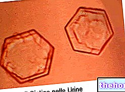
The urinary sediment is given by the "set of microscopic debris, cellular and otherwise, which can be found in the urine, in varying concentrations depending on the state of health of the patient. The examination of these sediments, using the microscope or recently introduced automated techniques, represents an integral part of traditional urine tests, capable of providing useful indications for the diagnosis of numerous pathologies.
Depending on the needs, in the urinary sediment it is possible to search and quantify the presence of blood cells, such as red and white blood cells, epithelial cells, microorganisms, etc. The urine sample is taken in the morning, because at this time the urine is more acidic and concentrated, thus allowing easier identification of the cellular elements and cylinders. We then proceed with the centrifugation and the eventual coloring; the important thing is that the urine is fresh, to avoid increases in pH and loss of cellular and organized elements.

The urine of healthy individuals contains a very limited number of red blood cells (red blood cells), leukocytes (white blood cells) and cylinders. In relation to the morphology and the qualitative-quantitative aspects of these elements, the examination of the urinary sediment can provide useful indications for the diagnosis of important pathologies, such as urethritis, prostatitis, balanitis, cystitis, kidney stones (lithiasis), glomerulonephritis, candidiasis, nephropathy diabetic, nephrotic syndrome, neoplasms, heavy metal intoxication, systemic lupus erythematosus, parasitosis, liver damage and many other diseases related to changes in the urinary sediment.


