What is Defecography?
Defecography is a radiological examination used in suspected or overt cases of obstructed constipation.
The purpose of the procedure is to identify any abnormalities of the anus and rectum, from a morphological and functional point of view.
Synonyms
Defecography is also called cinedefecografia or evacuative proctography. The examination called cystocolpography also involves the opacification of the urinary bladder and vagina (see below).

What is obstructed constipation?
Also called dyschezia, constipation due to obstructed defecation is a particular form of constipation in which the transit of stool is slowed down due to a rectal problem.
The most common causes include:
- morphological changes of the rectum: rectal prolapse, rectocele, mucosal invaginations;
- functional causes: paradoxical contraction of the puborectal muscle (which contracts instead of relaxing when defecating) or other dysfunctions of the perineum.
Functional causes can be the consequence of various factors:
- poor physical activity;
- weakness of the muscles of the perineum and abdominal wall;
- anal hypertonus;
- habit of postponing defecation for social, psychological reasons or because it causes pain (anism, anal fissures, etc.);
- chronic constipation;
- chronic abuse of irritating laxatives.
Purpose of the Examination and Execution
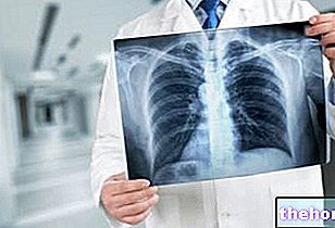
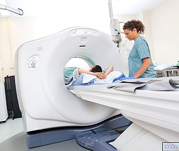
Defecography is a dynamic radiological examination: thanks to X-ray beams (or radio-magnetic fields), the doctor evaluates the way in which the patient expels a special contrast agent from the rectum introduced into the rectal area with a probe.
The introduction of this contrast medium into the rectal ampulla has the purpose of stretching its walls, causing an expulsion of the same which simulates natural defecation.
By observing the radiographic images, the doctor is able to detect any anatomical anomalies, such as prolapses, invaginations and rectoceles.
In addition to the morphological exploration, defecography allows to study the anorectum, and indirectly the pelvic floor, also from a functional point of view.
During the examination the patient is seated on a special radiolucent chair, equipped under the seat with a removable container which will collect the expelled material.
The collaboration of the patient is very important, as he will have to comply with the doctor's requests to contract, push and / or relax at specific times.
Variants
In women there may also be a need to evaluate the simultaneous presence of bladder or vaginal pathologies (cystocele, colpocele, etc.). In this case it is also necessary to opacify the bladder and / or the vagina with a different contrast medium.
If extended to the study of the bladder and vagina, the examination is more correctly defined as cystocolpodecography (or perineography).
Defecography can also include the intake of a contrast medium by mouth in order to study the possible presence of enterocele (bowel prolapse). In this case the examination times are considerably prolonged, as it is necessary to wait at least one "now to allow time for the contrast medium to reach and distribute in the intestine."
After the exam
The patient will be able to continue to emit clear stools in the following days.This is due to the gradual elimination of the bariate contrast medium injected into the rectum and possibly taken by mouth during defecography.
In the case of cystocolpodefecografia, the urine following the examination may show traces of blood; this is due to the trauma caused by the introduction of the catheter necessary to inject the contrast medium into the bladder. The same catheter, albeit rarely, can also cause urinary infections.
Preparation
A cleansing enema is usually required at least three hours before the exam. This prevents formed stools from interfering with the visualization of the anorectal morphology. The hospital will provide specific instructions to the patient on how to prepare for defecography.
Before proceeding with the actual defecography, the patient may be asked to sit on a radiological table for the acquisition of preliminary images in the supine position.
Precautions and Risks
Precautions
Since it is an examination that exposes to ionizing radiation, it must be avoided in cases where it is not possible to exclude a pregnancy in progress. This problem does not arise in the case of adopting the most modern magnetic resonance defecography.
In general, it is not necessary to interrupt any ongoing drug therapies.
Risks
Defecography is a particularly safe procedure, but like all invasive tests it can be burdened with possible complications. Although rarely, the contrast medium can develop a "local inflammation. Even more rare cases of intestinal perforation caused by the injection of air and contrast medium; this risk becomes more concrete in the presence of chronic inflammatory conditions of the intestine, such as Crohn's disease or ulcerative colitis.

