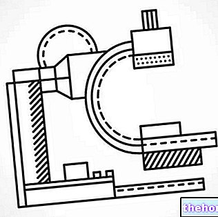
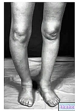
The production of images is possible thanks to a particular technological tool, which emits ionizing radiation.
As regards the realization of a chest X-ray, this occurs in a very simple way: the patient is positioned between the instrument that emits ionizing radiation (behind) and the photographic plate or digital detector for recording the radiation (in front , in direct contact with the chest).
Once the instrument has been activated, the outgoing radiations hit the chest of the individual under examination and, depending on how they are absorbed by the various anatomical structures, are impressed on the plate with different shades. For example, bones appear white because they absorb a lot of radiation, while the lungs appear black because they absorb little radiation.
Generally, the examination is carried out standing up, but, in certain situations, it can also be performed lying down, on a bed specially designed for this purpose.
and possibly also severe and / or persistent cough, chest pain, chest pain from trauma, fever.
Thanks to the reproductions that a chest X-ray is able to provide, doctors can analyze:
Lungs. Chest x-ray allows to diagnose various morbid conditions, including: pulmonary infections, cystic fibrosis, lung carcinomas, pulmonary emphysema, pneumothorax, etc.
The heart. Any heart abnormalities or malformations can be identified, such as valve defects or a condition called cardiac tamponade.
The blood vessels that branch off from the heart. Defects can be seen in the vessels that connect the heart to the lungs or in the vessels that connect the heart to various parts of the body (aorta).
The presence of calcium deposits in the blood vessels.
The presence of bone fractures.
Changes in the heart or lungs after surgery.
Placement of pacemakers, implantable defibrillators, or cardiac catheters.
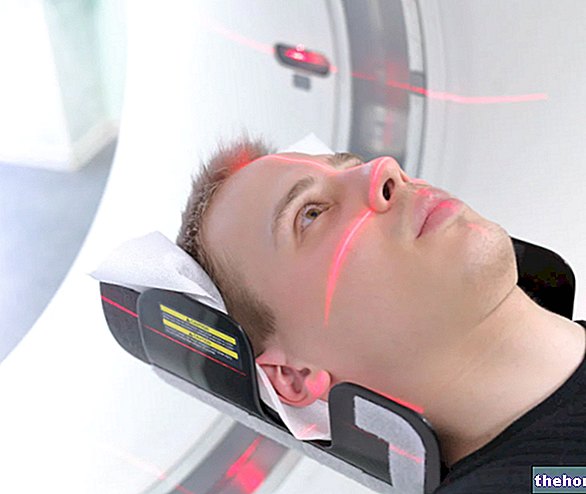
thoracic, produce internal images of the body, thanks to equipment that emits different doses of ionizing radiation.
But how is radioactivity measured and what is the precise amount of ionizing radiation that hits the patient during these tests?
First, the most commonly used unit of measurement for quantifying radioactivity is the millisievert (mSv).
Secondly, each radiological examination involves a specific "emission of ionizing radiation, which depends on the area of the body to be analyzed. For example, a chest x-ray is performed with a lower number of radiations than an x-ray of the abdomen, but higher than a dental x-ray and so on.
In addition to quantifying the radiation emitted by individual tests, the experts in the field have also tried to establish how many days / months / years of natural radioactivity it takes to develop the same radioactivity as a certain diagnostic test. Taking 3 mSv as an average reference value for natural radioactivity per year, the results that emerge are really interesting.
Continue reading Radiology Exams: Dental CT
-cos-come-si-svolge.jpg)
