Generality
Cornea transplantation, also known as keratoplasty, is the surgical operation of partial or total replacement of the cornea; this operation is used in cases of damaged or no longer functional cornea, to replace it with an analogue healthy, synthetic or taken from a recently deceased donor.

There are three types of corneal transplant: perforating (or penetrating) keratoplasty, lamellar keratoplasty and endothelial keratoplasty.
After the operation, the patient must follow some important medical instructions to avoid unpleasant complications.
A transplanted cornea can last up to 25 years.
What is corneal transplant?
Cornea transplantation, or keratoplasty, is the surgical intervention through which the operating doctor provides for the total or partial replacement of the original cornea, no longer functional and heavily damaged, with a similar healthy element, of synthetic origin or from a deceased donor recently.
WHAT IS THE CORNEA? A BRIEF REVIEW
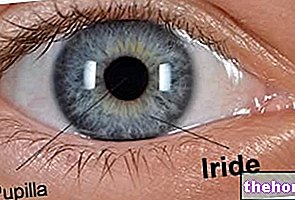
The cornea is the transparent, multilayered membrane that resides in the front of the eye and covers the iris and pupil.
Devoid of blood vessels (therefore not vascularized), this particular membrane represents the first "lens" that light encounters when it reaches the eye.
The functions of the cornea, which deserve a particular mention, are at least three:
- Protection and support of ocular structures;
- Filtration of some ultraviolet wavelengths → the cornea allows light rays to pass through the eye tissue, rather than being reflected or absorbed.
- Refraction of light → the cornea is responsible for 65-75% of the eye's ability to make the light rays coming from the outside converge on the fovea, ie the central region of the retina.

Uses
The cornea is a very delicate area of the eye with very little self-repair capacity.
This explains why an injury to him makes transplantation essential.
The most common medical conditions that require cornea transplantation are:
- Keratoconus: is, by far, the main cause of corneal transplantation;
- Degenerative diseases of the corneal tissue;
- Corneal perforation;
- Corneal infections that do not respond to any antibiotic treatment;
- The presence, on the cornea, of scars.
Procedure
Eye surgeons can perform cornea transplant surgery in at least three ways, the name of which is:
- Perforating keratoplasty or penetrating keratoplasty;
- Lamellar keratoplasty;
- Endothelial keratoplasty.
While perforating keratoplasty involves the replacement of an entire thickness of cornea and represents the oldest operating modality, lamellar keratoplasty and endothelial keratoplasty provide for the replacement of some layers of cornea and represent the most modern methods of intervention.
PERFORATING OR PENETRATING KERATOPLASTY
For perforating keratoplasty, the operating physician uses a kind of utility knife, called trephine, through which it cuts the damaged cornea section, for its entire thickness.
After resection, he removes the damaged corneal section and replaces it with the "new" one, synthetic or taken from a donor.
The grafting of the "new" cornea requires the application of several sutures, the removal of which, in some situations, can take place even 12 months after the procedure.
Perforating keratoplasty surgery can take place under general anesthesia or local anesthesia: in the first case, the patient is unconscious and insensitive to pain for the entire duration of the operation; in the second case, however, he remains conscious during the operation, but nevertheless does not feel any pain.
A classic perforating keratoplasty operation takes 45-60 minutes, including anesthesia.
As a rule, hospitalization is provided for one night, to allow the patient to recover better from the anesthesia and the very first effects of the surgical procedure.
LAMELLAR KERATOPLASTY
Through lamellar keratoplasty, eye surgeons transplant the outermost and possibly central layers of the cornea.
The instrumentation used may consist of the aforementioned trephine or in a particular laser, designed for the purpose.
The resection of the damaged corneal layers is followed by the application of healthy corneal layers, taken from a donor or of synthetic origin.
The grafting of the healthy layers of the cornea requires the realization of several sutures, exactly as in the case of penetrating keratoplasty.
There are two subtypes of lamellar keratoplasty:
- Anterior lamellar keratoplasty: consists in the removal and replacement of the outermost layers of the cornea.
- Deep anterior lamellar keratoplasty: consists in the removal and replacement of the outermost and central layers of the cornea.
At the end of the lamellar keratoplasty procedures, the patient can return home after a few hours from the conclusion of the operation, as long as the conditions are stable.
ENDOTHELIAL KERATOPLASTY
Through endothelial keratoplasty, eye surgeons transplant the innermost layers of the cornea and possibly the so-called corneal stroma (N.B: an anatomical description of the cornea is present here).
As in the case of lamellar keratoplasty, the tools used may consist of the usual trephine or in a laser beam designed for the purpose.
After resection of the damaged corneal layers, the application of healthy corneal layers, taken from a donor or of synthetic origin, follows.
For the grafting of the healthy layers of the cornea, sutures are not needed, but an air bubble, created specifically to keep the corneal transplant in place. This bubble resorbs autonomously within a few days, the time necessary for the graft to permanently attach itself to the rest of the cornea.
There are two subtypes of endothelial keratoplasty:
- Endothelial keratoplasty with stripping (or stripping) of Descemet's membrane: it consists in the replacement of the innermost layers of the cornea and 20% of the corneal stroma.
- Endothelial keratoplasty of Descemet's membrane: consists only in the replacement of the innermost layers of the cornea.
At the end of the endothelial keratoplasty procedures, the patient can return home after a few hours from the conclusion of the operation, as long as the conditions are stable.
Post-operative phase
Immediately following the cornea transplant procedure, the patient:
- He must keep the protective bandage applied to defend the operated eye for at least a whole day;
- You may experience slight eye pain. It's normal;
- He may suffer from blurred vision. It's normal.
POST-OPERATIVE RECOMMENDATIONS
Once home, the patient should pay attention to:
- Don't rub your eyes;
- Do not exert excessive physical exertion and do not lift weights;
- Do not go to places that are dusty, polluted or where smoke circulates;
- Use sunglasses as long as the sun causes discomfort;
- Do not practice contact sports, until otherwise indicated by the doctor;
- Wear protective goggles during certain sports activities, even if several months have passed since the intervention;
- Do not over-wet the eye during baths and showers for at least a month;
- Do not resume driving, until otherwise indicated by the doctor;
- Protect the eye with a bandage for at least a few weeks.
Risks and complications
One of the most feared complications of corneal transplantation is rejection, or the exaggerated reaction, set in motion by the immune system, against the "new" implanted organ. Moreover, the immune system of a specific organism purposely serves to recognize and attack everything that is foreign to the organism itself.
Fairly common phenomenon - in fact it affects one in 5 transplant recipients (therefore 20% of patients) - corneal rejection manifests itself with various symptoms and signs, including:
- Blurred vision
- Eye redness;
- Sensitivity to light (photophobia);
- Pain in the operated eye.
In the presence of such symptoms, it is advisable to contact your doctor as soon as possible, to expose the problem to him. With a timely measure, it is possible to stop the evolution of the complication.
The risk of rejection is increased by ocular inflammations, due for example to smoky environments, irritants, dust or particularly windy days.
OTHER COMPLICATIONS
In addition to rejection, corneal transplantation can also lead to other complications, such as:
- Astigmatism;
- Glaucoma;
- Uveitis;
- Retinal detachment;
- The reappearance of the morbid condition that made the transplant necessary;
- Small reopenings of surgical wounds. Remember that corneal repair is very slow, so a wound damaged by it heals very slowly;
- Infections, especially when surgical wounds are healing.
Results
The vision of an individual undergoing corneal transplantation can stabilize within a few weeks, as well as after a year or more.
The timing is affected by several factors, including: the mode of intervention and the condition of the cornea at the time of the operation.
Typically, if all goes well, transplanted corneas retain their transparency for about 25 years.
Readers can consult some of the most frequently asked questions related to cornea transplantation by clicking here.

