What is acute pyelonephritis
Acute pyelonephritis is a "rapid onset" infection of the renal pelvis and interstitial tissue of the kidney that generally affects young women.

The disease requires immediate intervention: if not treated properly, acute pyelonephritis can cause permanent damage to the organ and bacteria can spread to the bloodstream causing an "infection spread throughout the body."
Treatment of acute pyelonephritis includes antibiotic therapy and often requires hospitalization.
Diagnosis
The diagnosis of acute pyelonephritis is not always simple: there are differences in the clinical presentation and severity of the disease, in fact there is no coherent set of signs and symptoms that allows the pathology to be identified in a specific way (the symptoms could also be linked to other infections urinary tract, such as cystitis or urethritis).
In the outpatient setting, making the diagnosis of acute pyelonephritis usually begins with the collection of information about the patient's medical history, medical history and physical examination, and is confirmed by the results of urinalysis, which should include microscopic analyzes. Other laboratory investigations are used to identify the onset of secondary complications. Imaging studies are generally used in the following cases: suspected subclinical presentation of the disease, disease with atypical or insidious onset (gradual and usually associated with poor prognosis), resistance to therapy, the need to rapidly diagnose the onset of serious secondary complications (kidney stones, obstructive uropathy, perirenal abscess, etc.).
For this variety of reasons, physicians must maintain a high index of suspicion.
The presence of a symptomatology, characteristic of an infectious process, can guide the diagnosis:
CLEAR symptoms, indicative of acute pyelonephritis
High fever, lumbar pain, dysuria and renal involvement on physical examination.
Some Symptoms That May Cause DIAGNOSTIC UNCERTAINTY
The onset of kidney infection is sometimes manifested in the child only with the onset of fever, but is often associated with inappetence, abdominal pain, asthenia and foul-smelling urine. In the elderly patient, the only symptom may be a vague feeling of malaise. .
The microbiological investigation (microbiological urine culture + direct microscopic examination) allows to confirm the clinical suspicion in all these cases.
Physical examination
Doctors may suspect that a "kidney infection is in progress by performing a complete physical exam. The evaluation includes checking for clinical parameters, such as: heart rate, blood pressure, temperature control, and any signs of dehydration. The patient has Acute pyelonephritis commonly presents with lower back pain (in one or both kidneys), manifested by a "marked sensitivity of the kidney to palpation. If the affected person is a young woman, a pelvic exam may also be useful.
Laboratory investigations
Urinalysis: direct microscopy and microbiological culture
Microbiological diagnosis is a fundamental tool for providing a direct diagnosis.
Urine is the typical sample in which the causative agent of acute pyelonephritis is sought and must be analyzed by microscopy and culture, even in the case of a poor correlation between symptoms and bacteriuria. Urine culture must also be part of the "screening" of high-risk patients, such as pregnant women, the elderly, patients with catheters, subjects with anatomical-functional alterations in the urinary tract and in all cases of sepsis of unknown origin. . We also remember that the presence of bacteria in the urine (bacteriuria) can be "asymptomatic" and cause recurrence of the infection.
In order to obtain reliable results, the urine sample must be collected BEFORE THE BEGINNING OF THE ANTIBIOTIC THERAPY, appropriately, in order not to suffer any contamination: in carrying out the collection, by means of the intermediate cap technique, catheterization or suprapubic puncture, it must be taken in consideration of the presence of bacterial flora residing in the urethra and adjacent areas.
Direct microscopy
The direct microscopic examination allows to analyze a drop of fresh urine, then left to dry and processed with the Gram method (it allows to distinguish Gram-positive bacteria, which retain the basic dye assuming a violet color, from Gram-negative) .
The analysis of the urinary sediment allows to highlight if there is a condition of pyuria (presence of purulent material in the urine), as well as allowing the eventual identification of leukocytes and their quantification (leukocyte count).
Rapid urine test: dipstick
The test is done by dipping the test strips directly into the urine sample.
The dipstick allows you to quickly carry out some specific enzymatic tests, in order to highlight the enzymatic activity of leukocytes (esterases) and bacteria (nitrate-reductase, catalase, glucose-oxidase).
The examination allows to test the sample for some parameters relevant to the diagnosis of acute pyelonephritis:
- Presence of nitrites, from the transformation of nitrates carried out by pathogenic germs (if positive, it depends on the presence of an "adequate microbial load).
- Leukocyte esterase (confirms the presence of white blood cells). A positive result indicates a possible urinary tract infection.
- Hematuria and proteinuria, in acute pyelonephritis are parameters present in modest quantities, but indicative of the presence of blood and proteins in the urine.
Cultural examination
The urine sample is diluted and sown on culture media suitable for the growth of bacterial species which, more frequently, cause the onset of pyelonephritis; the procedure aims to determine the bacterial load (CFU / ml). L " standard urine culture is aimed at detecting non-fastidious microorganisms, such as enterobacteria, Gram-negative bacteria, Gram-positive bacteria, Staphylococcus spp., Streptococcus spp. and yeasts. On the other hand, specific microbiological analyzes make it possible to identify pathogens such as mycobacteria, anaerobic bacteria, etc. A bacteriuria that results significant from the culture test, must be evaluated according to various conditions and interpreted according to the individual case.
In front of a positive urine culture, the antibiogram is associated, which allows to evaluate the sensitivity of the pathogens, which intervene in the infection, to the various antibiotics.
The culture examination of the urine therefore assumes great importance, as it allows the isolation of the microorganism that causes the onset of acute pyelonephritis, confirms the diagnosis and facilitates the choice of adequate therapy based on the characteristics of the identified pathogen.
Visual exam
In acute pyelonephritis, urine is thick cloudy, due to the presence of purulent material.
The look opaque sample can be determined by the presence of erythrocytes, leukocytes, bacteria, epithelial cells or amorphous material.
Other evidence may support the findings:
- Antibody research: agglutination reaction for the detection of antibodies to enterobacteria. The presence of secretory type A (IgA) immunoglobulins indicates a local response and current or recent infection.
- PAR test (determination of the Residual Antibacterial Power): search for substances with antibacterial activity (usually certain drugs or chemotherapy).
Blood chemistry test
- Blood culture. Positive in about 12-20% of patients with pyelonephritis.
- Complete blood count, with complete blood cell count and with particular interest in detecting neutrophilic leukocytosis, typical of acute inflammatory processes.
- Inflammatory markers: presence of C reactive protein, high erythrocyte sedimentation rate (ESR).
- Procalcitonin. Recent studies identify it as a biological marker in the diagnosis of acute pyelonephritis in children under the age of two.
Farley test
The test is noteworthy as it is still present in scientific literature, however it is little used today as it requires a demanding maneuver with the introduction of a Farley catheter into an already infected urinary system:
- A urine sample is taken via catheter and cultured.
- The bladder is then emptied and treated with a solution containing an antibiotic and fibrinolytic enzymes.
- This solution is left in the bladder for 30 minutes to allow the elimination of the microbial load, before being emptied and washed with sterile saline.
- The physiological solution is eliminated from the bladder and 3 samples are taken, according to an interval of 10, 20 and 30 minutes.
If the infection affects the kidney, all the samples will be positive with a progressive increase in the titre (the bacterial load will be present in the first sample taken, as in all the following ones).
Imaging
Diagnostic imaging is useful in case of evidence of the clinical picture, to confirm the diagnostic suspicion or the presence of structural problems. Imaging is mandatory in patients with recurrent pyelonephritis and can help identify any obstructions (example: stones or strictures).
Spiral computed tomography (CT) is the best investigation in adult patients and can be used to confirm the diagnosis. Spiral CT uses no contrast media and reveals a moderate to severe pathological condition (as milder cases may be "normal").
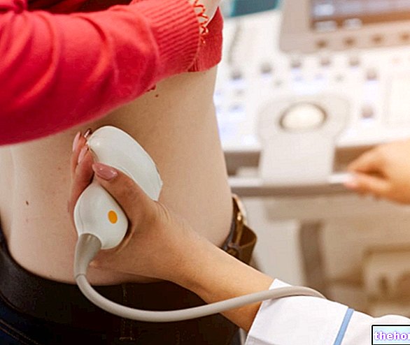
The ultrasound examination allows to identify abscesses, kidney stones or strictures.
For children, the choice may be between an ultrasound and computed tomography: CT is more sensitive, but the former is the safest option for the little patient (there is no radiation exposure).
Currently, magnetic resonance imaging (MRI) is still a "limited investigation in the evaluation of acute pyelonephritis, in terms of cost and availability. In adults, MRI can detect kidney infection, urinary tract obstruction, scarring and allow for evaluation of renal vascularity. In addition, magnetic resonance imaging, in the case of perirenal abscess, allows us to better define the extent of pyelonephritis than computed tomography.
Renal scintigraphy with 99mTc-DMSA (radiopharmaceutical consisting of technetium + dimercaptosuccinic acid, which is localized in the renal cortex) allows to detect anatomical and functional abnormalities of the kidneys in acute pyelonephritis (example: scars, distribution of real function, foci of infection ...).
Kidney biopsy
Renal biopsy identifies histological evidence of acute pyelonephritis and is occasionally used to rule out capillary necrosis or renal abscess formation.


-cos-cause-sintomi-e-rimedi.jpg)


.jpg)


