Generality
Keratoacanthoma is a benign skin tumor, which gives rise to a solid and isolated bump with a very characteristic appearance.

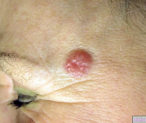
Figure: characteristic appearance of keratoacanthoma. From the site: dermapics.com
The neoplasm originates from a hair follicle or a pilo-sebaceous gland, develops within 6 weeks and then disappears within a few months.
The possible triggering causes are more than one: at the origin, in fact, there may be an excessive exposure to ultraviolet radiation, contact with certain toxic substances, a weak immune system (immunosuppression) and so on.
For a correct diagnosis, a physical examination at a dermatologist and a biopsy are required.
The therapeutic treatment adopted can be of a surgical or pharmacological type. The choice of a particular method of treatment depends solely on the characteristics of the keratoacanthoma.
What is keratoacanthoma?
Keratoacanthoma is a benign skin tumor that originates in a hair follicle or pilo-sebaceous gland. Due to its morphology and initial growth rate, it is very reminiscent of a malignant neoplasm of the skin, known as squamous cell carcinoma or squamous cell carcinoma; however, unlike this malignant tumor, keratoacanthoma is characterized by a spontaneous resolution (after about 5-6 months) and by a zero or almost no metastatic potential.
N.B: by metastatic power s "means the ability of the tumor to form metastases. Metastases are tumor cells that have moved from their original location and moved elsewhere, first contaminating the lymph nodes and then the other organs of the body.
THE PILIFEROUS FOLLICLE
The hair follicle is located in the dermis (layer of skin between the epidermis and the hypodermis) and is the structure within which a hair forms and grows.
The number of hair follicles on the skin is enormous and affects almost the entire human body; the areas that are totally devoid of it are the so-called hairless regions, such as the palm of the hand, the soles of the feet, the distal phalanx of the fingers, the lips, the glans and the clitoris.
Each hair follicle corresponds to a gland that produces sebum, also called the pilo-sebaceous gland.

Figure: overview of the internal anatomy of the skin. It is possible to recognize the layers of the skin (epidermis, dermis and hypodermis), the hair follicle and the pilo-sebaceous gland.
The growth of part of the body hair depends on the level of sex hormones circulating in the organism: androgens, that is the male sex hormones, stimulate its growth, while estrogens, that is the female sex hormones, have the opposite effect.
WHAT IS A BENIGN CANCER?
In medicine, with the term tumor, s "identifies a mass of very active cells, capable of dividing and growing in an uncontrolled way due to a genetic mutation of the DNA.
In a benign tumor, generally, the growth of the cell mass is very slow, it is not infiltrative (that is, it does not invade the surrounding tissues) and neither does it metastasize.
In other words, it is the exact opposite of a malignant tumor, which grows quickly and, if not removed in time from where it formed, can spread to surrounding tissues and the rest of the body.
EPIDEMIOLOGY
According to some English estimates, keratoacanthoma has an "annual incidence equal to one case in 1,000 people.
It usually affects individuals over the age of 60 (juvenile cases, in fact, are very rare) and is at least twice as frequent among males than among females.
In dark-skinned people (for example, Africans), it is very uncommon.
Causes
It has been shown that the formation of a keratoacanthoma can be caused by ultraviolet radiation (or UV rays) from the sun and tanning lamps, by contact with certain toxic substances (or chemical carcinogens), by a weak immune system (immunosuppression), by a physical trauma or a "pathogenic infection by a particular papilloma virus.
WHO IS MOST AT RISK?
After years of scientific and statistical studies, several conditions have been identified that favor the appearance of a keratoacanthoma. These risk factors are as follows:
- Have fair skin. Anyone can get keratoacanthoma, regardless of complexion. However, those with less melanin, the skin pigment that defends the skin from harmful UV rays, are more predisposed than those with more.
- Too much sun. Excessive exposure to sunlight, especially in the central hours of the hottest days, favors the onset not only of keratoacanthoma, but also of malignant skin tumors.
- Excessive use of tanning lamps. Tanning lamps emit the same ultraviolet radiation as the sun, so excessive use can have the same effects as prolonged sun exposure.
- Chronic immunosuppression. An individual's immune system is their defensive barrier against infections and other threats brought from the external environment. Conditions that create chronic weakening of the immune system (immunosuppression) favor the onset of various disorders, including keratoacanthoma and malignant skin tumors. An emblematic case of what has just been said is represented by leukemia or lymphoma patients and organ transplant recipients, who - being forced to suppress their immune system with special drugs - expose themselves to infectious diseases and, precisely, to malignant and benign skin tumors.
- Excessive exposure to tar and bitumen. According to some studies, bitumen and tar would contain carcinogenic chemicals, capable of promoting the appearance of keratoacanthoma. In fact, among the workers who treat these preparations almost every day, the incidence of this benign tumor is higher than normal.
- Infections induced by a particular strain of papilloma virus. The papilloma virus that induces the formation of warts seems to be involved, statistical data in hand, in the onset of keratoacanthoma.
- The advanced age and the male sex. The age of greatest onset is around 60, while the sex most affected is the male.
Comparing squamous cell carcinoma and keratoacanthoma, we can see the remarkable similarity that there is, from the etiological point of view (ie as regards the causes), between these two tumors, although the first is malignant and the second is benign.
Symptoms and Complications
Keratoacanthoma, in most cases, manifests itself with a skin mark that resembles a volcano. In fact, on the skin, a protuberance (or papule) appears with a small central crater filled with a particular protein of the cells of the epidermis, called keratin.
The areas usually most affected are the areas of skin most exposed to the sun, therefore: face, scalp, back of hands, ears, neck and legs (in women in particular).
The shape of the protuberance is rounded, the consistency is rigid and the color is identical to that of the skin or tending to red.
The dimensions may vary, depending on the patient in question, from a minimum of one centimeter to a maximum of 2.5 centimeters.
The growth rate is fast only in the first 2-6 weeks, after which it slows down until it almost disappears.
After about 5-6 months, this type of keratoacanthoma tends to disappear spontaneously, leaving, however, a noticeable scar.
OTHER TYPES OF KERATOACANTOMA
There is another type of keratoacanthoma, much rarer than the previous one, which causes an "itchy rash, characterized by many small papules and capable of affecting a" skin area of 5-15 centimeters. The papules differ from the more common type only in size, as they maintain the same volcano shape, the same keratin-filled crater, the same consistency and the same possibility of resolving spontaneously (but leaving a scar) after 5-6 months approximately.
This variant is also called generalized eruptive keratoacanthoma (in English, the acronym is GEKA) by Grzybowski, who was the first doctor to describe its characteristics.
WHEN SHOULD YOU WORRY ABOUT A KERATOACANTOMA?
Keratoacanthoma, as mentioned previously, has almost zero metastasizing power, so much so that it is considered, in effect, a benign tumor.
Nevertheless, those waiting for treatment are recommended to monitor the papule (s) on a daily basis, and to immediately contact their doctor if they notice a change in shape, color and / or size.
Diagnosis
To diagnose keratoacanthoma, we first proceed with an objective examination, during which the doctor analyzes the skin sign; subsequently, a biopsy is performed.
OBJECTIVE EXAMINATION
During the physical examination, the dermatologist thoroughly analyzes the appearance of the papule or papules, makes sure that there are no others elsewhere and, finally, traces the patient's clinical history, asking him certain questions. clinical is very important, because it allows us to understand if an individual falls into one of the risk situations described above.
Attention: the physical examination alone does not allow to establish the nature of the papule. In fact, it could be a keratoacanthoma, but also the result of an actinic keratosis or a malignant skin tumor, such as squamous cell carcinoma or basal cell carcinoma. That's why a more invasive diagnostic test, such as a biopsy, is needed.
BIOPSY
Biopsy is the only diagnostic test capable of detecting the exact nature of the papule that appeared on the skin. It involves the removal, through an "incision made on the suspected area, of a small portion of skin tissue, and the observation of this under the microscope. At the instrument, the cells of a keratoacanthoma have distinctive characteristics, very different, for example, from those of a malignant tumor.
Warning: the superficial biopsy of a keratoacanthoma does not allow to distinguish the latter from a malignant skin tumor, such as squamous cell carcinoma. Therefore, for a certain and precise diagnosis, a much deeper and more invasive incision and sampling are required.
Treatment
The most appropriate therapeutic treatment for keratoacanthoma is chosen based on the type of keratoacanthoma itself.
When keratoacanthoma presents with only one papule (the most common form), the ideal therapy is surgical removal of the bump.
When, on the other hand, it manifests itself with more papules (Grzybowski's generalized eruptive keratoacanthoma), a non-surgical approach must be adopted, based on local and systemic drugs; in these cases, surgery would be too invasive.
SURGERY
The most commonly used surgical methods to remove single papule keratoacanthoma from the skin are the following:
- Curettage and electrodissication. This intervention involves, first of all, the scraping (or curettage) of the superficial part of the benign tumor; subsequently, we proceed with the burning (electrodissecation) of the base of the keratoacanthoma. The scraping and burning are carried out, respectively, with a tool called "curette"and with an electric needle.
Not recommended for papules that form on the face, curettage and electrodissication represent an "excellent solution to not very large bumps located on the legs. - Excision or excision. It is the removal of the tumor area by incision. It is a moderately invasive operation and at risk of scarring, as the surgeon, to be sure of totally eliminating the papule, must also cut a part of neighboring healthy tissue. To close the "incision, sutures are applied.
- Mohs surgery. It consists in the elimination of the papule in small layers. Each layer, after removal, is observed under the microscope, the first of which without abnormal cells is the signal that the benign tumor has been completely removed.
Mohs surgery aims at the exclusive removal of the keratoacanthoma and the preservation of the underlying healthy tissues. It is an ideal method for papules formed on the nose, ears, lips and back of the hands. - Cryotherapy. It is cold therapy ("crio" comes from the Greek and means "cold"). It involves the use of liquid nitrogen, which, once applied to the keratoacanthoma, freezes and kills the cells of the tumor mass. Cryotherapy is suitable for not very large papules.
Sometimes, these treatments may end with a short course of radiotherapy, which serves to eliminate the last traces of keratoacanthoma.
NON-SURGICAL TREATMENT
Non-surgical treatment involves the use of local (ie directly on the affected area) and systemic (ie made to reach the affected area through the bloodstream) administration of anticancer drugs.
The most used preparations are:
- Retinoids, such as isotretinoin.
Method of administration: systemic, therefore the drug can be injected intravenously or taken by mouth.
- Methotrexate or methotrexate.
Method of administration: intralesional injection (N: B: for intralesional injection s "means that the drug is injected directly where c" is the keratoacanthoma).
- The 5-fluorauracil.
Method of administration: intralesional injection and local use.
- Bleomycin.
Method of administration: intralesional injection.
- Steroids.
Method of administration: intralesional injection.
- Imiquimod.
Method of administration: local use.
The non-surgical approach is adopted in Grzybowski's generalized eruptive keratoacanthomas, which cannot be removed surgically.
IN CASE OF NON-TREATMENT
Keratoacanthoma can resolve spontaneously, without any type of therapeutic treatment, within 5-6 months. However, this entails, as mentioned, the appearance of a visible scar.
Prognosis
Both surgery and the non-surgical approach provide good results.
However, at the end of the operation, it is recommended to undergo periodic checks at your dermatologist, in order to constantly monitor the progress of the situation. In fact, in some cases, keratoacanthoma can recur in the same spot and with the same characteristics.
Prevention
It is possible to prevent the formation of a keratoacanthoma by adopting the following recommendations:
- Avoid excessive sun exposure, especially if you have fair skin or are at high risk.
- Do not overuse the use of tanning lamps. Individuals at risk are strongly advised not to use them.
- Use sun protection creams, especially when you are at the sea or exposed to the sun in the central hours of the hottest days.
- If you are at risk of keratocanthoma, wear sunglasses and opaque clothing to protect the parts usually most exposed to sunlight.
- Check your skin periodically. It is good to examine, from time to time, the whole body, even the most unthinkable points.
- Don't overlook any skin abnormalities that appear suddenly and for no reason.
The above advice is also valid for preventing malignant skin tumors, the formation of which can have far more serious effects than a "simple" keratoacanthoma.


