Keyword
Functions of the immune system; primary and secondary lymphatic (or lymphoid) organs; White blood cells; antigens; macrophages; neutrophils; natural killer; dendritic cells; complement system; interferons; humoral immunity; cell-mediated immunity; antibodies; B lymphocytes; T lymphocytes; major histocompatibility complex.
agedTaken as a whole, the immune system represents a complex integrated network consisting of three essential components that contribute to immunity:
- the organs
- the cells
- chemical mediators
Read also: Natural Supplements to Strengthen Immune Defenses
- organs located in different parts of the body (spleen, thymus, lymph nodes, tonsils, appendix) and lymphatic tissues. They are distinguished:
- primary lymphatic organs (the bone marrow and, in the case of T lymphocytes, the thymus) are the site where leukocytes (white blood cells) develop and mature.
- secondary lymphatic organs capture the antigen and represent the site where lymphocytes can meet and interact with it; in fact they show a reticular architecture that traps foreign material present in the blood (spleen), in the lymph (lymph nodes), in the air (tonsils and adenoids) and in food and water (vermiform appendix and Peyer's plaques in the intestine).
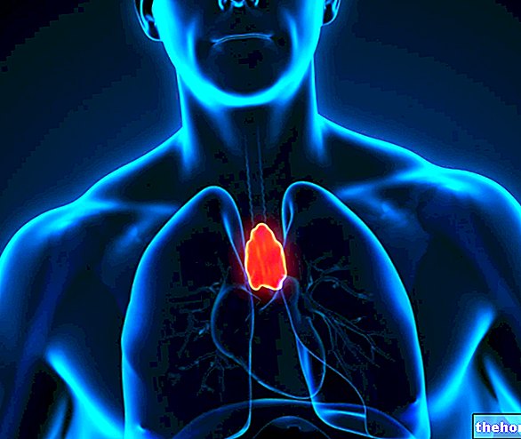
Deepening: the lymph nodes they play a very important role in the processing of the immune response, since they are able to trap and destroy bacteria and malignant tumor cells carried by the lymphatic vessels along which they are distributed.
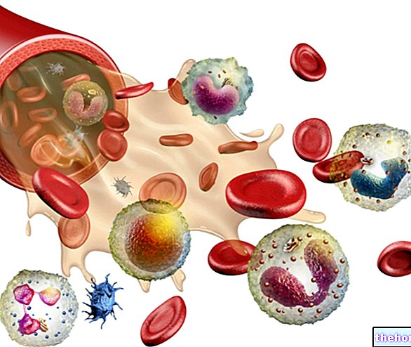
- isolated cells present in blood and tissues: the main ones are called white blood cells or leukocytes, of which different subpopulations are recognized (eosinophils, basophils / mast cells, neutrophils, monocytes / macrophages, lymphocytes / plasma cells and dendritic cells).
- chemicals that coordinate and carry out immune responses: through these molecules, the cells of the immune system are able to interact by exchanging signals that mutually regulate their level of activity; this interaction is allowed by specific recognition receptors and by the secretion of substances, generically known as cytokines, which act as regulatory signals.
The very important protective activity of the immune system is exercised through a triple defensive line which guarantees immunity, that is ability to defend against the aggression of viruses, bacteria and other pathogenic entities, to counteract damage or disease.
- Mechanical and Chemical Barriers
- Innate or Aspecific Immunity
- Acquired or Specific Immunity
The acid pH of sweat, conferred by the presence of lactic acid, associated with a small amount of antibodies, has an "effective antimicrobial action.
Enzyme present in tears, nasal secretions and saliva, capable of destroying the cell membrane of bacteria.
The oil produced by the sebaceous glands of the skin exerts a protective action on the skin itself, increasing its impermeability and exerting a mild antibacterial action (enhanced by the acid pH of sweat).
Viscous, whitish substance, secreted by the mucous membranes of the digestive, respiratory, urinary and genital systems. It protects us from microorganisms by incorporating them and masking the cellular receptors with which they interact to exercise their pathogenic activity.
It is able to fix and retain foreign bodies, filtering the air. Furthermore, it facilitates the expulsion of phlegm and of the microorganisms incorporated in it.
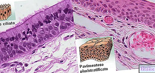
Cold viruses exploit the inhibitory action of cold on the motility of these cilia to infect the upper respiratory tract.
They prevent the proliferation of pathogenic bacterial strains by subtracting their nourishment, occupying the possible adhesion sites to the intestinal walls and producing active antibiotic substances that inhibit their replication.
Under normal conditions there is a saprophytic bacterial flora in the vagina which, together with the slightly acidic pH, prevents the excessive growth of pathogenic germs.
The normal temperature inhibits the growth of some pathogens, which is even more hindered in the presence of fever, which also favors the intervention of immune cells.
- Neutrophils
- Basophils
- Eosinophils
- Lymphocytes
- B lymphocytes
- Humoral immunity (antibodies)
- T lymphocytes
- Cell-Mediated Immunity
- B lymphocytes
Note: many texts include physical and chemical barriers within the innate immunity, we have treated them separately to give a better overview of the immune system.
It should be noted immediately that both types of immune responses are closely interrelated and coordinated; the innate response, for example, is reinforced by the acquired antigen-specific response, which increases its "efficacy. Overall" the resulting immune response proceeds according to the following basic steps:
- PHASE OF RECOGNITION OF THE ANTIGEN: identification and identification of the foreign substance
- ACTIVATION PHASE: communication of the danger to other immune cells; recruitment of other immune system actors and coordination of overall immune activity
- EFFECTIVE PHASE: attack on the invader with destruction or suppression of the pathogen.
The concept of antigen: the very functionality of the immune system implies the ability to distinguish harmless cells from dangerous ones, sparing the former and attacking the latter. There distinction between the self (or self) and the non-self (or non-self), between harmless and dangerous, is allowed by the recognition of particular surface macromolecules, called antigens, which have a unique and well-defined structure. For example, as we have seen, the innate immune system is able to recognize the lipopolysaccharide structure of the outer wall of bacteria.
Let's now look at some important definitions.
- Antigens are substances recognized as foreign (not self) and therefore able to induce an immune response and interact with the immune system.
- The epitope is the specific portion of an antigen, recognized by the antibody.
- The hapten is a small antigen capable of inducing an immune response only if conjugated to a carrier.
- The allergen is an element foreign to the organism itself non-pathogenic, but still capable of causing allergic disease in some individuals as a consequence of the induction of an immune response; examples are dust mites, pollen and molds.
- Autoantibodies are anomalous antibodies directed against the self, or against one or more substances of the organism; they are a fundamental element of autoimmune diseases, including rheumatoid arthritis, multiple sclerosis and systemic lupus erythematosus.
Present since birth and therefore called innate, non-specific immunity does NOT have any kind of memory towards previous encounters with pathogens. Furthermore, it is NOT strengthened following new and further contacts with the same pathogen.
As soon as the microorganisms manage to overcome the mechanical-chemical barriers, the non-specific immunity is activated RAPIDLY and helps to neutralize them, blocking many infections and preventing their evolution into disease. This ability is linked to the presence:
- on the one hand of particular cells, such as neutrophil granulocytes and monocytes;
- on the other of some particular substances produced by them that attract other cells of the immune system.
1) CELLULAR FACTORS
THE CELLS OF INNATE IMMUNITY
- Phagocytes, or Macrophages and Neutrophils: Phagocyte debris / pathogens.
- Natural Killer: Affect virus infected cells and cancer cells.
- Dendritic cells: present the antigen (APC cells) by activating cytotoxic T lymphocytes
- Eosinophils: They act on parasites.
- Basophils: Similar to Mast Cells; involved in inflammatory and allergic reactions.
- Phagocytes: recognize invaders through specific surface receptors, englobe them and destroy them by digesting them in lysosomes (phagocytosis); in addition, they attract other cells of the immune system by secreting cytokines.
The main phagocytes are tissue macrophages and neutrophils.- Macrophages: endowed with marked phagocytic activity, derive from monocytes produced in the bone marrow and circulating in the blood. They are present in all tissues and particularly concentrated in those most exposed to possible infections, such as the pulmonary alveoli. Neutrophils, on the other hand, circulate in the blood and only penetrate infected tissues.
In addition to phagocytic activity, in response to the presence of bacteria, macrophages secrete soluble proteins, called cytokines, chemical mediators that recruit other cells of the immune system:- Chemotaxins: attract other phagocytes, some stimulate the proliferation of B and T lymphocytes, others produce drowsiness
- Prostaglandins: produce the increase in body temperature to a level intolerable to pathogens and which stimulates the defenses: FEVER.
- Neutrophil granulocytes or leukocytes (polymorph) nucleated (PMN): they are blood cells capable of exiting the vessels to migrate into the tissues where the infection occurred and engulf, destroying them, microorganisms, debris and cancerous cells. They are capable of acting even in conditions they die at the site of infection, forming pus.
- Macrophages: endowed with marked phagocytic activity, derive from monocytes produced in the bone marrow and circulating in the blood. They are present in all tissues and particularly concentrated in those most exposed to possible infections, such as the pulmonary alveoli. Neutrophils, on the other hand, circulate in the blood and only penetrate infected tissues.
- NK lymphocytes - Synonyms: natural Killer (NK) cells): T lymphocytes are thus defined which, once activated, emit substances capable of neutralizing virus-infected and tumor cells. Stimulated by certain cytokines, natural killer lymphocytes cause virus-infected or abnormal cells to "commit suicide" by a mechanism known as apoptosis.
NK lymphocytes also have the ability to secrete various antiviral cytokines, including interferons.
Unlike the other types of lymphocytes (B and T), characteristic of the acquired immune response, the NK lymphocytes do not specifically recognize the antigen (they do not have specific receptors) and therefore are part of the innate immunity. - Dendritic cells: unlike macrophages and neutrophils, they are not able to phagocytize the antigen, but they capture it and expose it on their surface following the interaction with it (for this reason they belong to the group of APC cells, presenting the " antigen) In this way, the externalized antigen is recognized by the "killer" cells, the cytotoxic T lymphocytes that initiate the specific immune response. Not surprisingly, the dendritic cells are concentrated at the level of those tissues that act as a barrier with the external environment, such as the skin and the inner lining of the nose, lungs, stomach and intestines.
PLEASE NOTE: after having played the role of "sentinels" (intercepting the antigens and exposing them on their surface), the dendritic cells migrate to the lymph nodes where the T lymphocytes meet.
PLEASE NOTE:
The cells of innate immunity constitutively express multiple receptors on their surface, each of which recognizes more than one well-defined microbial structure, hence their capacity for multiple non-specific recognition.
2) HUMORAL FACTORS
- Complement system: plasma proteins produced by the liver, normally present in an inactive form; they are similar to messengers that synchronize communications between the various components of the immune system. Cytokines circulate in the blood and are sequentially activated, with a cascade mechanism (the activation of one triggers that of the others), in the presence of appropriate stimuli.
When activated, cytokines trigger a series of enzymatic chain reactions that make certain components of the immune system acquire particular characteristics. For example, they attract phagocytes and B and T lymphocytes to the site of infection via a mechanism called chemotaxis. The complement system also has an intrinsic ability to damage the membranes of pathogens by causing pores on them that lead to lysis. Finally, the complement covers the bacterial cells "tagging" them (opsonization) as a pathogen, facilitating the action of phagocytes (macrophages and neutrophils) which recognize and destroy them.
The opsonins are macromolecules which, if they cover a microorganism, enormously increase the efficiency of phagocytosis as they are recognized by receptors expressed on the phagocyte membrane. In addition to the opsonins deriving from complement activation (the best known is C3b), one of the most powerful opsonization systems is represented by the specific antibodies that cover the microorganism and which are recognized by the phagocyte Fc receptor. Antibodies (or immunoglobulins) represent the humoral defense mechanism of acquired immunity.
PLEASE NOTE: complement activation is a mechanism common to both innate and acquired immunity. There are in fact three distinct pathways of complement activation: 1) the classical pathway, mediated by antibodies (specific immunity); 2) the alternative pathway, activated directly by some proteins of the cell membranes of microbes (innate immunity); 3) the lectin pathway (it uses mannose as a site of attachment to the membranes of pathogens).
- Interferon System (IFN): Cytokines produced by NK lymphocytes and other cell types, so named for their ability to interfere with viral reproduction. Interferons facilitate the intervention of cells that participate in the immune defense and inflammatory reaction.
There are various types of interferon (IFN-α IFN-β IFN-γ), produced by some T lymphocytes after the recognition of an antigen. Interferons are active against viruses, but do not attack them directly, but stimulate other cells to resist them; particularly:- they act on cells not yet infected inducing a state of resistance to viral attack (interferon alpha and interferon beta);
- they help activate Natural killer (NK) cells;
- stimulate macrophages to kill tumor cells or cells infected with viruses (gamma interferon);
- inhibit the growth of some cancer cells.
- Interleukins: act as "short-range" chemical messengers, acting especially between adjacent cells:
- Factors of tumor necrosis: secreted by macrophages and T lymphocytes in response to the action of the interleukins IL-1 and IL-6; they allow to raise the body temperature, dilate the blood vessels and increase the catabolic rate.
Inflammation is a characteristic reaction of innate immunity, very important for fighting infection in damaged tissue:
- attracts substances and immune cells to the site of infection;
- produces a physical barrier that delays the spread of the infection;
- when the infection is resolved, it promotes repair processes of the damaged tissue.
The inflammatory response is triggered by the so-called degranulation of mast cells, cells present in the connective tissue which, following the insult, release histamine and other chemicals, which increase blood flow and capillary permeability and stimulate the intervention of white blood cells. Typical symptoms of inflammation are redness, pain, warmth and swelling of the inflamed area.
PLEASE NOTE: in addition to infections, the inflammatory response can also be triggered by stings, burns, injuries and other stimuli that damage tissues.
The main cellular actors of the immune system involved in inflammation are neutrophils and macrophages.
, in particular towards some very specific molecules (antigens) of the pathogen.
The acquired immunity is strengthened following further contacts with the same pathogen (memory appearance of the recognition carried out).
Acquired immunity intervenes only when the other lines of defense have not been able to effectively counter the pathogen. It overlaps the innate immunity by strengthening the immune response: inflammatory cytokines attract lymphocytes to the site of the immune reaction and the latter then release their own cytokines fueling and enhancing the specific inflammatory response.
There are two types of acquired immune response:
- humoral (or antibody-mediated) immunity: it is mediated by B lymphocytes that transform into plasma cells that synthesize and secrete antibodies
- cell mediated (or cell mediated): mediated mainly by T lymphocytes that directly attack the invading antigen (intervention of helper and cyto-toxic T lymphocytes)
Acquired humoral immunity can also be divided into active (it is the organism itself that produces antibodies in response to exposure to pathogens) and passive (antibodies are acquired from another organism, for example from the mother during fetal life or by vaccination).
1) HUMORAL FACTORS
- Immunoglobulins (antibodies): some microorganisms have developed tricks to alter their surface markers, becoming "invisible" to the eyes of phagocytes and losing the ability to activate complement. To combat these pathogens, the immune system produces specific antibodies against them, labeling them as dangerous to the eyes of phagocytes (opsonization). The antibodies coat the antigens facilitating their recognition and phagocytosis by the immune cells. The function of antibodies is therefore to transform unrecognizable particles into "food" for phagocytes.
Antibodies are part of the globulins (globular plasma proteins) present in the blood and are called immunoglobulins. They are cataloged in 5 classes, namely: IgA, IgD, IgE, IgG and IgM.Antibodies can also bind and inactivate some bacterial toxins and help fuel inflammation by activating complement and mast cells.
Immunogenic antigens are molecules capable of stimulating the synthesis of antibodies; in particular, all these molecules have a small part able to bind to its specific antibody. This portion, called the epitope, generally differs from antigen to antigen. It follows that each antibody recognizes and is sensitive only to one or more specific epitopes and not to the whole antigen.
2) CELLULAR FACTORS
The cells mainly involved in the establishment of acquired immunity are the antigen presenting cells (the so-called APC, antigen-presenting cells) and the lymphocytes.
LYMPHOCYTES
- B and T lymphocytes: B lymphocytes originate and mature in the bone marrow, while T lymphocytes originate in the bone marrow, but migrate and mature in the thymus. As we have seen, these organs are called primary lymphoid organs and, in addition to production, they are also responsible for the maturation of these lymphocytes.
During its development, each lymphocyte synthesizes a type of membrane receptor that can only bind to a certain antigen. The link between antigen and receptor therefore gives rise to the activation of the lymphocyte, which at that point begins to divide repeatedly; in this way lymphocytes are formed with receptors identical to the one that had recognized the antigen: these lymphocytes are called CLONES and the process with which they are formed is called CLONAL SELECTION.
PLEASE NOTE: following the activation of the lymphocytes, both EFFECTIVE CELLS are formed, which will actively participate in the immune response, and MEMORY CELLS, which have the task of recognizing the antigen in the event of a subsequent invasion.- EFFECTIVE CELLS: ready to face the enemy and destroy him
- MEMORY CELLS: they do not attack the foreign agent but enter a state of quiescence ready to intervene in a subsequent attack OF THE SAME IDENTICAL ANTIGEN
B Lymphocytes express immunogobulins (Antibodies, Ab), while T Lymphocytes express receptors; both act as membrane receptors. - B LYMPHOCYTES: they directly recognize the antigen through surface antibodies; once activated they partially proliferate and mature in specialized cells that secrete antibodies (called plasma cells, real "antibody factories") and partly in cells of the memory (which have the same function as the previous ones but are longer-lived and for this reason they continue to circulate for much longer periods than plasma cells, sometimes even for the entire life of the organism). As we have seen, memory cells ensure rapid production of antibodies should a certain pathogen reappear for the second time.
Each B lymphocyte expresses on its membrane something like 150,000 identical and specific antibodies (receptors) for the same antigen. The antigen-antibody binding is extremely specific: there is an antibody for every possible antigen. A mature plasma cell can produce up to 30,000 antibody molecules per second.
PLEASE NOTE: activation of B lymphocytes requires stimulation of T helper lymphocytes. B lymphocytes recognize antigen in native form, while T lymphocytes recognize ancillary cell-processed antigen (APC)
- T LYMPHOCYTES: they interact directly with the cells of our body that are infected or altered. They contribute to the elimination of the antigen:
- directly, cytotoxic activity towards virus infected cells;
- indirectly, by activating B lymphocytes or macrophages.
- THE T helper lymphocytes they govern the regulation of all immune responses by releasing cytokines that help cytotoxic B lymphocytes and T lymphocytes. They therefore have a COORDINATION FUNCTION:
- have CD4 membrane receptors;
- recognize antigens presented by MHC II;
- they induce differentiation of B lymphocytes into plasma cells (the latter producing antibodies);
- they regulate the activity of cytotoxic T lymphocytes;
- activate macrophages;
- they secrete cytokines (interleukins);
- there are several subtypes of helper T lymphocytes; for example, Th1s are important in the control of intracellular pathogenic bacteria through the activation of macrophages.
- THE cytotoxic T lymphocytes (TC) (CD8 +) govern the cell-mediated immune response and exert a "toxic action against their specific target cells (infected cells and cancer cells). They therefore have a function of DEMOLITION OF FOREIGN CELLS:
- they present the membrane molecule CD8;
- recognize the antigens presented by MHC I;
- they selectively target virus-infected and cancer-causing cells;
- regulated by T Helper.
When an infection has been defeated, the activity of the B and T lymphocytes is blocked thanks to the action of other T lymphocytes called suppressors which, in fact, suppress the immune response: however, this process is not entirely clear and is currently a source of several studies
PLEASE NOTE: B lymphocytes recognize antigens in soluble phase, while T lymphocytes cannot bind to antigens unless they exhibit MHC class I protein sequences on their cell membranes. T lymphocytes therefore recognize antigens presented by "APCs. "(antigen presenting cells).
The tools of the acquired immune system to recognize specific antigens are therefore three:
- Immunoglobulins or Antibodies
- T cell receptors
- Major histocompatibility complex and MHC proteins on APC (antigen presenting cells).
Molecular complexes (fragments of antigen + MHC II molecules) are exposed on the surface of some cells, which are therefore called antigen presenting cells (APC). APC cells (dendritic cells, macrophages and B lymphocytes) can be compared a of the shuttles that present on the cell surface protein fragments derived from the digestion of proteins internalized by phagocytes combined with the major histocompatibility complex of class 2.
At this point it is necessary to specify that there are two types of MHC molecules:
- MHC class I molecules are found on the surface of almost all nucleated cells and ensure that the "abnormal" body cells are recognized by the CD8 receptors of cytotoxic T lymphocytes; it is therefore possible to "avoid a massacre" that is to prevent the cytotoxic lymphocytes from attacking the healthy cells of the organism. For example, natural killer lymphocytes recognize how non-self cells with low expression of MHC-I (tumor cells), while cytotoxic T lymphocytes attack only cells that have viral antigen complexes - MHC-I.
- MHC class II molecules, on the other hand, are found only on APC cells of the immune system, mainly on macrophages, B lymphocytes and dendritic cells. Class II MHCs exhibit exogenous peptides (derived from the digestion of the antigen) and are recognized by the CD4 receptors of T helper lymphocytes.
The peptides exposed on the cell surface thanks to the MHC are passed to the screening of the cells of the immune system, which intervene only if they recognize these complexes as "not self".
After the exposure of the antigen-MHC complex, the cells migrate through the lymphatic vessels to the lymph nodes, where they activate other protagonists of the immune system; in particular:
- If a cytotoxic T cell encounters a target cell that exposes fragments of antigen on its MHC-I (tumor nucleated or virus infected cells) it kills it to prevent reproduction;
- If a helper T cell encounters a target cell that exposes exogenous antigen fragments on its MHC-II (phagocytes and dendritic cells) it secretes cytokines increasing the immune response (eg by activating the macrophage or antigen-presenting B lymphocyte).







.jpg)
