Generality
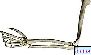
The bones of the hand are, in the human being, the skeletal structure of the terminal tract of each upper limb. They are a total of 27 and, according to the anatomists, can be divided into three large groups: the carpal bones (or carpal or carpal bones) , the metacarpal bones (or metacarpals) and the bones of the fingers of the hand (or phalanges of the hand).
The carpal bones represent the proximal portion of the skeleton of the hand; the metacarpals represent the intermediate portion of the skeleton of the hand; finally, the phalanges of the hand represent the distal portion of the skeleton of the hand.
The bones of the hand contribute to the grip capacity of the hand, guarantee stability to the child while walking on all fours, constitute very important joints (eg: the wrist joint) and, finally, insert the tendons of the hand muscles.
Like any bone in the human skeleton, the bones of the hand can also fracture.

What are the bones of the hand?
The bones of the hand are, in the human being, what constitutes the skeleton of the terminal part of each upper limb.
Inside the human body, the hands are two anatomical structures that serve to:
- Grasping objects;
- Perceiving through the sense of touch;
- To communicate;
- To guarantee stability, during the first years of life, when the human being still walks on all fours.
Anatomy
In all 27, the bones of the hand can be divided into three large groups: the carpal bones (or more simply carpus), the metacarpal bones (or metacarpals) and the bones of the fingers of the hand (or phalanges of the hand).
The carpal bones are 8 and represent the proximal skeletal portion of the hand; the metacarpal bones are 5 and represent the intermediate skeletal portion of the hand; finally, the phalanges of the hand are 14 and represent the distal skeletal portion of the hand.
In anatomy, proximal and distal are two terms with opposite meanings.
Proximal means “closer to the center of the body” or “closer to the point of origin.” Referring to the femur, for example, it indicates the portion of this bone closest to the trunk.
Distal, on the other hand, means "farther from the center of the body" or "farther from the point of" origin. "Referred (again to the femur), for example, it indicates the portion of this bone farthest from the trunk (and closest to the "knee joint).
CARPUS BONES
Irregularly shaped, the 8 carpal bones form the anatomical region of the wrist in two rows: a proximal row, close to the radius arm and ulna bones, and a distal row, bordering the base of the metacarpal bones.
The bones of the proximal row are: scaphoid, lunate, triquetroum and pisiform.
The bones of the distal row, on the other hand, are: trapezius, trapezoid, capitate and hooked.
- Proximal carpal bones. The bones of the proximal row play a fundamental role in the constitution of the wrist joint (which should not be confused with the aforementioned anatomical region).
While the scaphoid and semilunar articulate with the two articular surfaces of the radius, the triquetroum and pisiform inserts an important ligament that comes from the styloid process of the ulna. - Distal carpal bones. The carpal bones of the distal row have the important task of articulating the carpus to the metacarpals.
While the trapezius, trapezoid and capitate articulate with the base of one metacarpal bone only, the hook joins two adjacent metacarpal bones.
To be precise, the trapezius borders on the metacarpus that precedes the thumb; the trapezoid makes contact with the metacarpus that precedes the index; the capitate is at the base of the metacarpus that precedes the middle finger; finally, the hook is articulated with the metacarpals that precede the ring and little fingers.
METACARPAL BONES
The metacarpal bones, or metacarpals, are long bones, arranged parallel to each other, in which it is possible to distinguish three regions: a central region, called the body; a proximal region, called the base; finally, a distal region, identified with the term head.
The base of the metacarpals borders on the carpal bones according to the scheme described in the previous sub-chapter: therefore, starting from the side of the thumb, the base of the metacarpal preceding the thumb adheres to the trapezius; the base of the metacarpus that proceeds the index finger adheres to the trapezoid; the base of the metacarpus that precedes the ring finger adheres to the capitate; finally, the bases of the metacarpals that precede the ring and little fingers adhere to the hook.
The head of the metacarpals is the region that makes contact with the first phalanx of the fingers: it results that each metacarpus corresponds to a finger of the hand.
Between the base of the metacarpals and the carpal bones are a series of joints, as well as between the head of the metacarpals and the first phalanges of the hand.
HAND MUDLANGES
Cylindrical in shape, the phalanges of the hand are the skeleton of the 5 fingers of the hand.
Except the thumb - the only one formed by 2 phalanges - all the other fingers of the hand have 3 phalanges each.
The phalanges closest to the head of the metacarpals are called first phalanges (or proximal phalanges); starting from these, the following ones are called second phalanges (or intermediate phalanges) and third phalanges (or distal phalanges).
Between each phalanx there is a "joint", which gives the fingers of the hand a certain mobility.
In the case of arthrosis (or osteoarthritis), the joints present between the second and third phalanges of the fingers of the hand are the joint elements that develop the so-called Heberden's nodules.
Note: in the first finger of the hand, the numbering of the phalanges ends with the second phalanges.
Functions
The bones of the hand and their particular arrangement play a decisive role in some functions of the hand, such as in the grasping of objects or in the baby's walking on all fours.
Furthermore, the bones of the hand form very important joints (eg: the wrist joint), they insert the ligaments which are a fundamental part of the aforementioned joints and are the attachment point of the tendons belonging to the so-called muscles of the hand.
Pathologies
Like all bones in the body, the bones of the hand can also fracture.
There are three classes of fractures affecting the bones of the hand: fractures of the carpal bones, fractures of the metacarpals (or metacarpal fractures) and fractures of the phalanges.
FRACTURE OF A CARPUS BONE
The bones of the hand located in the carpus, which most commonly suffer a fracture, are the scaphoid, lunate and trapezius.
The main causes of scaphoid fractures include falls with the hands extended forward; typical causes of lunate fracture include direct blows to the wrist and chronic trauma; finally, the classic causes of trapezius fracture include violent blows to the back of the hand and falls with a hand closed in a fist and radial deviation (ie bent towards the radius).
The characteristic symptom of carpal bone fractures is pain.
For a correct diagnosis, X-ray examination is essential.
The therapy of composite fractures of the carpal bones involves the application of a cast on the patient's hand. The cast can last from a minimum of 4 to a maximum of 12 weeks.
Unlike the previous case, the treatment of displaced fractures of the carpal bones involves the use of surgery. In these circumstances, the purpose of surgery is to fix the various separated bone segments together by means of screws and pins.
Adequate treatment is essential to avoid long-term complications (e.g. wrist arthritis).
FRACTURE OF A METACARPUS
The bones of the hand with the metacarpal site, which fracture more easily, are the first metacarpus - precisely the base of the first metacarpal - and the fifth metacarpal - to be precise, the region just before the head.
In medicine, the fracture of the base of the first metacarpus is called a Bennett fracture, while the fracture of the region that precedes the head of the fifth metacarpus is called a boxer's fracture.
Typically, Bennett's fracture occurs after a "hyperabduction of the thumb. A boxer's fracture, on the other hand, is a consequence of punching objects with some resistance; it is called a boxer's fracture, because it is typical of boxers.
To diagnose metacarpal fractures, X-ray examination is essential.
Treatment of a metacarpal fracture depends on the severity of the injury.
In fact, if the fracture is stable and not particularly serious, doctors opt for the application, on the patient's hand, of a plaster splint, to be kept in position for about 2-3 weeks. If the fracture is stable and severe, it is a cast of the affected hand is expected for at least 4-6 weeks. Finally, if the fracture is unstable, the therapy chosen by the doctors is surgical and consists of an intervention aimed at joining, by means of screws, the separated bone portions.
FRACTURE OF A PHALANGE OF THE HAND
Fractures of one or more phalanges of the hand are conditions of mild severity, which arise as a result of traumatic events to the damage of the fingers of the hand (eg: crushing of a finger). In general, the treatment of fractures affecting the bones of the hand making up the fingers simply consists of a period of rest.



.jpg)


