Anatomy
The duodenum is the first portion of the small intestine, a long canal that extends from the pylorus (final part of the stomach) to the ileocecal sphincter (initial section of the large intestine), dividing into three portions: duodenum, jejunum and ileum.

If upstream we find the stomach with its pylorus, downstream of the duodenum we find the jejunum, from which it separates by means of the duodenal-jejunal fissure.
Of all the segments of the small intestine, with its 25-30 centimeters, the duodenum represents the shortest tract, but also the most important from the digestive point of view; it is no coincidence that the word duodenum means "twelve fingers" corresponding precisely to about 25 centimeters. In addition to being particularly short, this portion of the small intestine is also quite large (average caliber: 47 mm) and fixed, given its close adherence to the posterior abdominal wall. Morphologically, the duodenum has the shape of a C, with the convexity on the right and the concavity, where the head of the pancreas is located, located on the left.
Didactically, the duodenum is divided into four portions: superior or bulb, descending, horizontal and ascending.

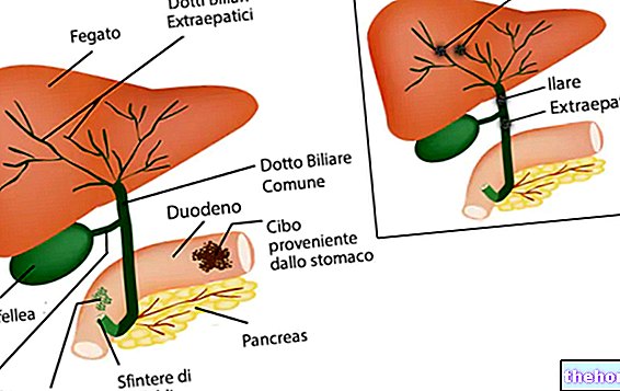
The descending portion, or second part of the duodenum, runs along the right side of the vertebral column and the inferior vena cava. It represents the direct continuation of the upper tract and continues with the horizontal part through the right duodenal flexure. This portion receives the secretion of the liver and pancreas: the bile carried by the choledochus and the pancreatic juice from the "duct of the same name, converge for a very short distance before flowing into the lumen of the duodenum, about 7-10 centimeters from the pylorus, in a dilation called papilla of the Vater, in whose outlet there is a particular smooth muscle formation, known as sphincter of Oddi or greater duodenal papilla.The accessory pancreatic duct instead opens two centimeters higher, at the level of the minor duodenal papilla.

The third portion of the duodenum runs horizontally and, in the postero-superior region, is in close relationship with the head of the pancreas. Finally, the fourth and last portion of the duodenum, the ascending one, rises along the left margin of the aorta to the level of the second lumbar vertebra, where it turns sharply forward to continue in the jejunum, forming the duodenodijunal flexure.
Physiology of the duodenum
The digestive activity of the duodenum is quite intense, since it collects the secretion of very important glands, such as the liver (bile), the pancreas (pancreatic juice), those of Brunner (duodenal glands that secrete an alkaline mucus) and intestinal ones (juice enteric).
The digestive juices have the purpose of neutralizing the acidity of the gastric chyme and completing its digestion. In addition, the villi appear in the duodenum, characteristic of the entire small intestine and responsible for the absorption of nutrients (thanks to the cells of the brush border which cover them).
In addition to the digestive and absorbent function, the duodenum also has activities:
- motor: it is the seat of peristaltic movements designed to mix the food material with the digestive juices, making them progress along the intestine;
- endocrine: the duodenum secretes various hormones with endocrine and paracrine action, such as secretin, cholecystokinin, gastrin, GIP, VIP, somatostatin and others (all important for adapting digestive functions to the quantity and quality of the food contained in the digestive tract, but also the state of health of the organism);
- immune: the GALT lymphoid tissue present in the mucosa of the duodenum constitutes the first barrier against any pathogens.





.jpg)
