The bronchi represent the airways contiguous to the trachea, which - in the adult - bifurcates at the level of the 4th-5th thoracic vertebra to give rise to the two primary or main bronchi, one for the right lung and one for the left lung. primary bronchi are in turn subdivided into branches of ever smaller caliber, forming the so-called bronchial tree (just like a plant, they form branches that progressively decrease in size).

Like the upper airways (nasal cavities, nasopharynx, pharynx, larynx and trachea), the bronchi are essentially responsible for transporting air from the external environment to the functional units of the lungs, the alveoli, in which gas exchange takes place (the pulmonary alveoli are small air-filled sacs, densely surrounded by capillaries and responsible for the exchange of oxygen and carbon dioxide).
The structure of the primary bronchi is identical to that of the trachea; as such, they maintain a cartilage support structure in their wall. By gradually branching into ducts of lower caliber, the bronchi give rise to the so-called bronchioles, in which the cartilage structure described above is lost.

After penetrating into the relative lung lobes, each lobar or secondary bronchus is subdivided into the various bronchopulmonary segments. Inside the lungs, the lobar bronchi lose the cartilage support structure typical of the trachea and primary bronchi (C-rings), becoming covered with irregular plates of hyaline cartilage, while the smooth muscle forms complete rings (unlike what happens in the trachea, where the posterior cartilaginous openings are filled by the tracheal muscle.) In this way the intrapulmonary bronchi no longer have a part flattened at the back, but are completely rounded.

As one enters the bronchial tree, the thickness of the bronchial walls decreases together with the caliber of the airways, which are less and less rich in cartilage tissue and increasingly rich in muscle tissue.
As soon as they penetrate the lung lobes, the secondary bronchi subdivide into smaller branches, the so-called tertiary (or segmental) bronchi. Each of these branches out by serving with smaller branches distinct sections of lung tissue, called bronchopulmonary segments. As shown in the figure, each lung is in fact divided by 10 bronchopulmonary segments, separated from each other by connective tissue.
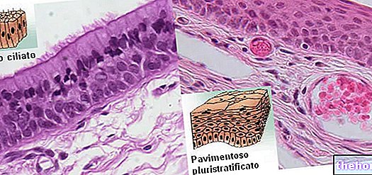
From the tertiary bronchi, through repeated ramifications, the so-called bronchioles originate. As anticipated, as the bronchial airways become thinner, the amount of cartilage in their wall also decreases; at the same time, the number of glands and goblet cells (important for preventing the entry of germs and dust) decreases, while the contribution of smooth muscle tissue and elastic tissue increases. Furthermore, the height of the epithelium progressively decreases, while in the terminal bronchioles the hair cells become cuboidal (from columnar or cylindrical), losing the cilia and flattening further in the areas responsible for gas exchange (where "muscle tissue is absent).
About 78,000
In turn, the bronchioles subdivide repeatedly giving rise to smaller and smaller ducts, the so-called terminal bronchioles, with a diameter of less than 0.5 mm. These form the terminal part of the conduction system of the respiratory system; in fact they supply the pulmonary acini with air where gas exchanges take place.

The bronchioles have neither glands nor cartilage in their wall, while they are equipped with a continuous layer of smooth muscle that provides support to the mucosa; they also contain the so-called Clara cells, which replace the mucipar goblet cells and are presumably responsible for protecting the respiratory epithelium from bacteria, toxins and collapse, also providing for its regeneration in case of damage.
Inferiorly, the terminal bronchioles continue with the respiratory bronchioles, which differ considerably from the progenitors in that they are provided with alveoli that open directly on their wall; therefore they have a dual function, both of conduction and of gas exchange.








.jpg)
