What is it and why is it done?
The blood smear is a laboratory test that allows to obtain a sort of photograph, a "snapshot of the cell population present in a drop of blood. Through the smear on the slide it is in fact possible to evaluate the number - but also the morphology, the stage of maturation and the numerical percentages - of red blood cells, leukocytes and platelets.

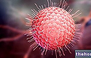
As shown in the figure, thanks to the blood smear it is also possible to highlight the presence of parasites, such as those responsible for malaria, heartworm and sleeping sickness. We are therefore talking about a precious diagnostic aid, a second level test which however requires a certain dexterity and experience on the part of the operator. Generally, the execution of the smear follows a first-line examination, such as the blood count, considered suspicious The complete blood count is an automated test that determines the number of blood cells; the smear, on the other hand, is a manual examination, which involves the observation and direct counting of these cells.

When taking the smear
Your doctor may prescribe a blood smear when:
CBC and differential leukocyte counts indicate a suspicion of abnormality or immature cells;
in the presence of a suspicion of a deficiency, a disease or a disorder in the production of blood cells;
you want to monitor the efficacy or side effects of a therapy (eg chemotherapy) or the evolution of a haematological disease (eg leukemia).
How is a blood smear done?
To make a smear
- place a drop of the patient's blood near the end of a special slide which will act as a base, suitably washed, degreased in alcohol and carefully dried.
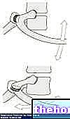
- near the end of the drop of blood which is furthest away from the corresponding side of the basal slide, place the shorter side of a second ground slide slightly smaller than the previous one at an angle of about 40 °
- pull the ground slide towards the drop, so that the latter spreads by capillarity along the contact line of the two slides.
- push the ground slide towards the opposite end, with constant, rapid and light movement, in order to obtain the smear;
- dry the smear, then proceed to the specific fixing and staining step
- a specialist (laboratory doctor) examines the blood smear thus obtained under a microscope, making a general assessment of the cell populations present, such as number, size, shape and general appearance. As shown in the image at the beginning of the article, the streaked drop contains thousands of erythrocytes, hundreds of leukocytes and numerous platelets (cell fragments involved in clotting). Red blood cells are the predominant cells by number.

Select Blood Tests Blood Tests Uric acid - uricaemia ACTH: adrenocortitotropic hormone Alanine amino transferase, ALT, SGPT Albumin Alcoholism Alphafetoprotein Alphafetoprotein in pregnancy Aldolase Amylase Ammonemia, ammonia in the blood Androstenedione Antibody-endomysial antibodies Anti-gliadicides Nucleus Helicobacter pylori antibodies Embryo carcinoal antigen - CEA Prostate specific antigen PSA Antithrombin III Haptoglobin AST - GOT or aspartate aminotransferase Azotemia Bilirubin (physiology) Direct, indirect and total bilirubin CA 125: tumor antigen 125 CA 15-3: tumor antigen 19-9 as tumor marker Calcemia Ceruloplasmin Cystatin C CK-MB - Creatine kinase MB Cholesterolemia Cholinesterase (pseudcholinesterase) Plasma concentration Creatine kinase Creatinine Creatinine Creatinine clearance Chromogranin A D-dimer Hematocrit Blood culture Hemocrome Hemoglobin Glycated hemoglobin a Blood tests Blood tests, Down syndrome screening Ferritin Rheumatoid factor Fibrin and its degradation products Fibrinogen Leukocyte formula Alkaline phosphatase (ALP) Fructosamine and glycated hemoglobin GGT - Gamma-gt Gastrinemia GCT Glycemia Red blood cells Granulocytes HE4 and Cancer at "Ova" Immunoglobulins INR Insulinemia Lactate dehydrogenase LDH Leukocytes - white blood cells Lymphocytes Lipases Tissue damage markers MCH MCHC MCV Metanephrines MPO - Myeloperoxidase Myoglobin Monocytes MPV - average platelet volume Natremia Neutrophils Homocysteine Thyroid hormones OGTT Osmocyte Plasma protein A associated with pregnancy Peptide C Pepsin and pepsinogen PCT - platelet or platelet hematocrit PDW - distribution width of platelet volumes Platelets Plateletpenia PLT - number of platelets in blood Preparation for blood tests Prist Test Total IgEk Protein C (PC) - Protein C Activated (PCA) C Reactive Protein Rast Protein Test Specific IgE Reticulocytes Renin Reuma-Test Oxygen Saturation Sideremia BAC, BAC TBG - Thyroxine Binding Globulin Prothrombin Time Partial Thromblopastin Time (PTT) Activated Partial Thromboplastin Time (aPTT) Testosterone Testosterone: free and bioavailable fraction Thyroglobulin Thyroxine in the blood - Total T4, free T4 Transaminases High transaminases Transglutaminase Transferrin - TIBC - TIBC - UIBC - saturation of transferrin Transtyretin Triglyceridemia Triiodothyronine in the blood - Total T3, free T3 Troponin TRH and Troponins of s thymol to TRH TSH - Thyrotropin Uremia Liver values ESR VDRL and TPHA: serological tests for syphlis Volemia Conversion of bilirubin from mg / dL to µmol / L Conversion of cholesterol and triglyceridemia from mg / dL to mmol / L Conversion of creatinine from mg / dL to µmol / L Conversion of blood glucose from mg / dL to mmol / L Conversion of testosteronemia from ng / dL - nmol / L Conversion of uricemia from mg / dL to mmol / L



.jpg)
