Generality
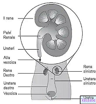
Urography is a radiological procedure that allows to evaluate the urinary system from a morphological and functional point of view. This clinical investigation, also known as intravenous pyelography, uses X-rays and a contrast agent to examine the kidneys, ureters, bladder and urethra.
Urography is indicated for diagnosing disorders affecting the urinary tract, such as stones, trauma, obstructions, birth defects or tumors.

Diagnostic value
The urography uses a contrast medium (generally an iodine solution) which allows to accurately evaluate the structures of the urinary tract. On radiographic images, after intravenous injection, this "dye" becomes visible almost immediately (appears bright white). The contrast agent is then removed from the bloodstream via the kidneys and urine produced by them. The radiograms, obtained during the intravenous urography, allow to clearly visualize the structures through which the contrast medium passes, allowing to examine the anatomy of the interested parts and to determine if they are performing their physiological function correctly.
The doctor may recommend intravenous urography if the patient has signs and symptoms that may be related to a urinary tract disorder (such as, for example, blood in the urine, back and flank pain, etc.). Intravenous urography, in fact, it can be used to diagnose a number of conditions affecting the urinary tract, such as:
- Kidney and bladder stones;
- Kidney cysts;
- Inflammation and infections of the bladder and kidney;
- Tumors of the urinary tract (example: renal cell carcinoma, transitional cell carcinoma, etc.);
- Obstruction (for example, at the level of the vesicoureteral junction);
- Anatomical abnormalities of the urinary tract;
- Enlarged prostate.
An intravenous urography can be done in both emergency and routine situations:
- Emergency. This procedure is performed on patients who present to the emergency room with symptoms suggestive of a severe obstructive urological condition, such as severe renal colic accompanied by hematuria. In this circumstance, the doctor may suspect a kidney stone, which is causing the urinary system to be obstructed. Patients are usually monitored for follow-up and further treatment. The radiographic images, obtained in a double time interval. "(30 minutes after injection, 1 hour, 2 hours, 4 hours, etc.), provide important information for the urologist on the location and severity of the obstruction.
- Routine. This procedure is more common for patients with "unexplained microscopic or gross hematuria. Urography is used to check for a tumor or similar disorder that alters the anatomy of the urinary tract."
Preparation
- A blood test may be required prior to the procedure to verify proper kidney function (creatinine, BUN, etc.). The kidneys must in fact be able to filter the contrast medium; therefore, intravenous urography is rarely done if you have kidney failure.
- The patient should inform the doctor if he has any allergies, especially to contrast media. Urography is also contraindicated in cases of iodine intolerance, severe heart disease, multiple myeloma and ongoing pregnancy.
- Before the procedure, the patient may be required not to eat for several hours and to take some laxatives for a day or two. This measure makes sure that the intestine is free of large amounts of stool, which can make interpretation of the radiographic images more difficult.
- In case of diabetes and metformin therapy it may be necessary to stop taking the drug 48 hours before and after the procedure, as an "interaction with the contrast agent" may occur (it is possible to discuss with your doctor the appropriate management diabetes during this period).
During urography
The sequence of images is roughly collected as follows:
- The patient lies supine on an examination table. A first frontal radiograph of the abdomen is taken. The contrast medium is then injected into a vein in the arm or hand. The dye begins to be excreted through the kidneys, ureters and bladder.
- During the next 30-60 minutes, at certain intervals of time (approximately every 5 minutes), radiograms of the kidney area are collected. Each time an X-ray is to be taken, the patient is asked to hold his breath. The radiographic images show the contrast medium as it travels through the urinary system at various stages Immediately after administration of the contrast medium it is filtered through the renal cortex At an interval of 3 minutes, the calyxes and renal pelvis are visible. At 9-13 minutes, the contrast begins to empty into the ureters and travel to the bladder, which begins to fill. About 10 minutes after the injection of the contrast medium, the doctor may eventually apply compression to the lower abdominal area, to cause the distention of the upper urinary tract and evaluate certain conditions (the maneuver is contraindicated in cases of obstruction). To visualize the bladder correctly, the patient is asked to empty the bladder before collecting the last X-ray image. The contrast medium collected in the organ, in fact, could mask a pathology.
After the urography
Intravenous urography is usually completed in 30-60 minutes. The patient should be able to return to normal activities as soon as the procedure is finished. It is advisable to drink plenty of fluids to help remove all of the contrast medium. body.
Results
During the urography, the following are evaluated:
- Kidneys: regular appearance, smooth contours, size, location, filtration and flow;
- Ureters: size, regular appearance and symmetry;
- Bladder: complete emptying, smooth and regular appearance.
A radiologist examines and interprets the radiographic images obtained during intravenous urography and sends a report to the physician, with whom the patient can discuss the results.
Side effects and contraindications
An intravenous urography is generally a safe diagnostic investigation and complications are rare.
- After injecting the contrast medium, it is possible to experience a metallic taste in the mouth and a tingling or warm sensation (similar to hot flushes). Some people experience a general feeling of discomfort, headache, nausea or vomiting. These side effects are almost always temporary.
- In a small number of cases, an allergic reaction to the contrast agent used in the test may occur. Symptoms may be mild (for example, itchy rash and mild swelling of the lips). More serious manifestations include: cardiac arrest and collapse due to extremely low blood pressure, difficulty breathing, and other symptoms of anaphylactic shock. It must be emphasized that severe reactions are rare and, should it be necessary, the hospital ward has access to full resuscitation equipment during the procedure.
- During intravenous urography, the patient is exposed to a very limited amount of radiation, so he is not susceptible to any harm. However, the investigation is contraindicated during pregnancy, as the fetus is more sensitive to the associated risks (malformations).
- Acute renal failure is a rare complication of intravenous urography. The contrast medium used during the procedure can cause damage in people with poor kidney function.
Final remarks
As some investigations, such as computed tomography and magnetic resonance imaging, take less time and provide more detail on the anatomy and function of the structures being evaluated, the clinical application of intravenous urography has become less common.
However, this can still be a valid diagnostic tool, particularly for:
- Identification of some structural disorders of the urinary tract;
- Kidney stone detection;
- Provide information on urinary tract obstruction.








.jpg)
