Edited by Dr. Stefano Casali
The chest wall
The coasts


The Diaphragm
The muscles of the diaphragm are divided into sternal, costal, which are inserted on the last six ribs, and vertebral, inserted on the arcuate ligaments and on the vertebral processes.
The Diaphragm
seen from below

Defects of the sternal muscles (foramen and Morgagni's hernia), of the muscles of the ligaments or of the posterior ribs (Bochdalek's hernia) are frequent, and there are frequent communications between the peritoneum and the diaphragm, more frequent on the right, which are at the base of the pleural effusions in course of subdiaphragmatic pathology (Meigs syndrome, peritoneal dialysis etc.).
The diaphragm is innervated by the phrenic nerves, which run into the mediastinum. The sensory innervation of the diaphragm is scarce. Moreover, the sensory fibers of the phrenic are localized at the shoulder level, so that diaphragmatic pain can be referred to the shoulder, and neuritis of the relative metamers can cause diaphragmatic paralysis.
The intercostal muscles
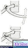
The external intercostal muscles run downwards and forwards, the internal intercostals downwards and backwards. A thin muscular layer is located immediately below the parietal pleura. The intercostal vessels and nerves run below and inside the lower edge of the rib (important for pleural puncture).
The chest wall and the pleural cavity


The airways
- The upper airways include:
the nasal cavities, the pharynx, the larynx
- The lower airways include:
the trachea, which originates from the cricoid cartilage, has a length of 10 11 cm and bifurcates at the height of the 5th vertebra; the main bronchi and their branches
The trachea and the bronchi
The trachea consists of a cartilage wall (15-20 rings linked anteriorly by a


Lobule and pulmonary acinus:
Towards the limit of each auriferous path, the terminal bronchiole is reached. The lobule represents the structural unit of the lung and is made up of three or five terminal bronchioles. Each lobule is made up of 10-15 elementary units, the pulmonary acini. , or respiratory unit, is defined as the part of the lung fed by a terminal bronchiole. The berries vary in size and shape; in the adult the grape can reach up to 1 cm in diameter. Inside the berry you can see from three to eight generations of respiratory bronchioles which have in part

Second part "









.jpg)
