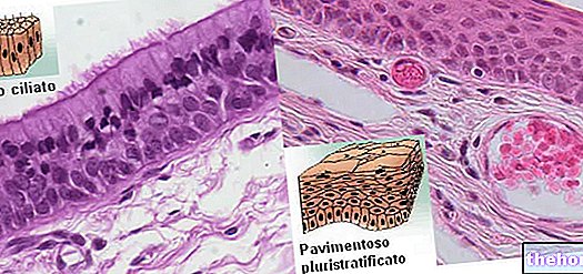
The intestinal mucosa is the innermost layer of the organ. As such, it directly faces the lumen of the intestine, in close contact with the products of digestion.
Below the mucosa, proceeding outwards, the remaining tunics meet: the submucosa, the muscularis and the serosa.
The intestinal mucosa is in turn made up of three distinct portions: a single layer of epithelial cells, a connective tissue that holds the epithelium (the lamina propria) in place and a small layer of smooth muscle, called muscularis mucosae, that separates it from the tunic below.
In the small intestine, the intestinal mucosa has a velvety appearance, conferred by the widespread presence of small finger-like ridges called intestinal villi.
Just as plants absorb nutrients into the soil through their roots, intestinal villi absorb substances essential for cell growth and survival.
The intestinal villi (see figure) have the purpose of increasing the absorbent surface, further extended by the lifting of the mucosa in macroscopic folds and by the sinking of the epithelial layer:
the numerous folds of the intestinal mucosa of the duodenum and jejunum are called folds, semilunar folds or conniving valves, while the tubular invaginations of the epithelium, which carry deep into the supporting connective tissue, are called crypts.

The particular conformation of the absorbent cells, also known as enterocytes, has the purpose of maximizing the digestive and absorption capacity of the organism.
Among the numerically most numerous enterocytes stand out some goblet cells that secrete mucus into the intestinal lumen. This viscous and lubricating substance is responsible for protecting the intestinal mucosa from the insults of acids and digestive products, as well as from digestive enzymes that could attack it.
In the furrows arranged between the villi we find a further component, represented by the Galeazzi's glands or Lieberkuhn's crypts, spread throughout the small intestine and also present in the large intestine. Crypts are made up of cells that will mature into enterocytes (absorbent cells), of others that process mucus, of serous cells that synthesize enzymatic proteins, of Paneth cells that produce lysozyme and other antimicrobial enzymes, and of silver-affine cells of the gastro-endocrine system. intestinal, which produce endocrine and paracrine hormones to facilitate digestion and absorption.
Intestinal mucosa "








.jpg)


