Cartilage: what it is and what it is for
Articular cartilage is an elastic tissue with considerable resistance to pressure and traction (it is a specialized connective with a supporting function). It has a pearly white color and covers the extremities of the articular bones protecting them from friction. Its function is similar to that of a shock absorber which with its action safeguards the normal joint relationships and allows movement.

The cartilage tissue is not vascularized as it is devoid of blood capillaries. Cartilage (with the exception of articular hyaline cartilage) is surrounded by a layer of dense connective tissue (perichondrium), rich in blood vessels, which allow it to feed by diffusion. Diffusion feeding of chondrocytes is a slow and much less effective process than blood circulation; for this reason the regenerative capacities of this tissue are very low.
In our body, three types of cartilage tissue with different characteristics and functions are commonly distinguished:
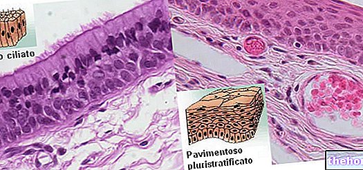
- hyaline cartilage: bluish-white in color is the most abundant type of cartilage. In the fetus it forms a large part of the skeleton and as it grows it is almost completely replaced by bone *. In the adult it forms the costal, nasal, tracheal, bronchial and laryngeal cartilages and covers the articular surfaces. The cartilage is covered by a thin envelope of compact connective tissue called the perichondrium. Near the articular surfaces this tissue disappears.
- elastic cartilage: opaque yellow in color, it has particular characteristics of elasticity. It constitutes the scaffolding of the auricle, of the epiglottis, of the Eustachian tube and of some laryngeal cartilages.
- fibrous cartilage: whitish in color, it is particularly resistant to mechanical stress. It is found at the insertion point of some tendons on the skeleton, in the intervertebral discs, in the menisci of some joints (knee) and in the pubic symphysis
* until the end of the growth between the epiphysis and the diaphysis of the long bones a small area remains called the epiphyseal disc which continues to proliferate cartilage tissue. This tissue is gradually transformed into bone ensuring normal skeletal elongation. With the attainment of maturity, the disc too it becomes ossified and the bone will no longer be able to grow.
Cartilage lesions
The strength and functionality of the cartilage tissue are exceptional. Suffice it to say that it normally resists almost 80 years of continuous stress and that no device built by man can boast the same properties.
However, during the life span this resistance can be undermined by a series of factors that expose the cartilage to more or less important lesions. Cartilage lesions are normally classified into two distinct categories:
primary or post-traumatic events that arise as a result of accidents of a mechanical nature (fractures, sprains, stress fractures) or are linked to genetic factors
secondary or degenerative that arise as a result of continuous stress or problems of a metabolic or immune nature (for example as a result of a deficit of the immune system such as in rheumatoid arthritis)
Regardless of its nature, a lesion of the articular cartilage marks the beginning of osteoarthritis.
Osteoarthritis is, by definition, a degenerative pathology of the articular cartilage. In Italy over 4 million people suffer from it, especially the elderly. More than 80% of people over the age of 55 show radiographic signs of osteoarthritis (especially women). The pain associated with it involves limitations in movement and represents a great cost to society. Knee, hands, hip and spine are the most affected sites.
Arthritis is a degenerative inflammatory disease that affects the joints. It manifests itself with inflammation, pain and stiffness in movements, up to deforming, in the most serious cases, the affected joints. There are various types of arthritis that arise for different reasons.
Patellar chondropathy (or chondromalacia) is quite frequent in athletes and in the long run can lead to knee arthrosis. The cause of origin is linked to the excessive stress to which the knee is subjected during sports activity. Then there are a whole series of predisposing factors (such as muscle and joint imbalances) that contribute to the premature onset or worsening of the disease. Even acute trauma, such as a fall can contribute to its onset.
Patellar chondropathy affects the protective cartilage layer behind the patella that wears down over time. In most cases it is asymptomatic but sometimes the subject complains of widespread pain around the patella associated with mild swelling (especially in more severe cases).
Prevention of cartilage injuries
Cartilage, although poorly vascularized, is a living tissue that responds to external stimuli. In particular, the proliferation and functionality of chondrocytes is regulated on the basis of the mechanical stress suffered by the joint. If these stimuli fail, as happens following prolonged immobility (fracture), the production of proteoglycans slows down. And it is precisely from this consideration that the importance of regular physical activity in the prevention of osteoarthritis can be deduced.
Exercise also helps improve mood and appearance, decreases pain, increases elasticity and keeps body weight under control, improving balance and decreasing the risk of falls.
The importance of physical exercise also derives from the consequent muscle strengthening. This last point plays an important role in the prevention and treatment of patellar chondropathy. The strengthening of the quadriceps and in particular of the vastus medialis is very important for patellar stabilization and of the knee joint in general. It is performed thanks to a tool called leg extension working in the last degrees of extension with the toes pointing towards the " external.
Diet also plays an important role in the prevention of cartilage lesions and if in the past someone tried to draw up a whole series of useful and harmful foods, today the general guideline is to propose a balanced and varied diet. The rules to follow are not specific for arthritic disease but general. It is therefore advisable to limit saturated fats, to prefer foods of biological origin, to take the right quantities of fiber, vitamins and minerals, as widely explained in the article: dietary advice.






.jpg)
