Generality
Rectal prolapse consists of the exit, through the anal canal, of a portion of the rectum. The exact causes are not yet known. However, it is suspected that there may be a general weakening of the pelvic muscles at the origin.

The symptoms are different and their appearance depends on the degree of severity of the rectal sliding. Severe rectal prolapse significantly affects the quality of life of those who suffer from it.
The possibilities for treatment are numerous. There are both conservative and surgical treatments. The choice of the therapeutic path and its success are based on several factors, such as: severity of rectal prolapse, associated diseases, age and general health of the patient.
Brief anatomical recall: the pelvic floor and the rectum
To understand what happens in rectal prolapse, it is advisable to do a brief anatomical review, concerning the pelvic floor and the rectum.
THE PELVIC FLOOR
The pelvic floor is the set of muscles, ligaments and connective tissue, located at the base of the abdominal cavity, in the so-called pelvic area. These structures cover a fundamental and indispensable function: they serve to support and maintain the urethra, the bladder in their positions. , the rectum and, in women, the uterus.

THE RECTUM INTESTINE
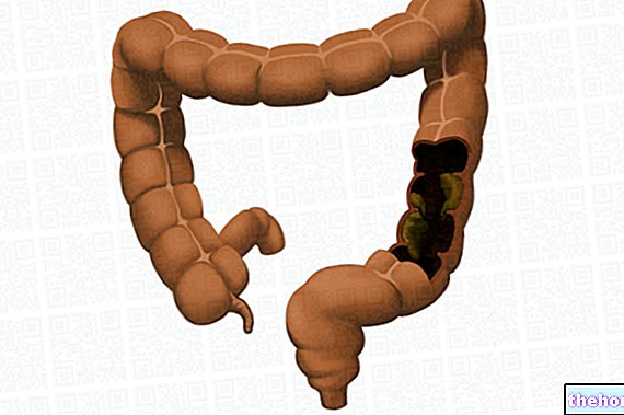
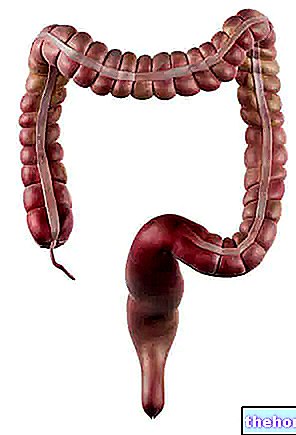
The rectum (or rectal canal) is the last portion of the intestinal canal. Approximately 13-15 cm long, it is placed between the sigma tract of the intestine and the anus (or anal canal). The walls of the rectal canal consist of three different layers of fabric:
- The mucosa, in direct contact with the lumen of the rectal canal
- A layer of muscle tissue
- A layer (on the outside) of adipose tissue, the mesorectum
The rectum is the point of collection of feces, before their evacuation; evacuation, which is controlled by the contraction of the muscles and ligaments of the pelvic floor.
What is rectal prolapse
Rectal prolapse is the sliding of the rectum downwards, with the escape of its internal walls, or only of its mucosa, through the anus.
CLASSIFICATION OF RECTAL PROLAPSE
Sometimes, rectal prolapse causes the walls, which make up the rectal canal, to protrude; in other cases, however, it causes only the mucosa to escape or an internal failure, not visible from the outside.
In light of this, the following types of rectal prolapse can be distinguished:
- Complete rectal prolapse. Characteristics: the walls, which constitute the rectal canal, completely protrude from the anus.
- Rectal prolapse of the rectal mucosa (or partial prolapse). Characteristics: the mucosa is the only part of the rectum that protrudes from the anus.
- Internal rectal intussusception. Characteristics: the rectum has slipped on itself, without, however, protruding from the anal canal.
This classification is the best known. However, it should be remembered that each type of rectal prolapse can be divided into further subtypes, different for some clinical characteristics. In order not to complicate this text, we have chosen to report only the three main categories.
EPIDEMIOLOGY
The exact data on the incidence of rectal prolapse is unknown. Certainly, the confirmed cases are less than the real ones.
The most affected subjects are adults, especially those of advanced age (over fifty) and women. However, rectal prolapse can also occur in some young individuals (rare) and in children between the ages of one and three.
Causes of rectal prolapse
The exact cause of rectal prolapse is not yet known. The most accepted hypothesis is that there is a weakening of the structures (muscles, ligaments and connective tissue) of the pelvic floor. Below we address the possible causes of this weakening.
RISK FACTORS
Several risk factors appear to be involved, which strain and traumatize the muscles, ligaments and connective tissue of the pelvic area.
- Increased abdominal pressure, due to:
- constipation
- diarrhea
- BPH
- pregnancy
- chronic bronchitis (for example, chronic obstructive pulmonary disease and cystic fibrosis)
- Previous pelvic organ surgery
- Parasitic infections (for example, amoebiasis and schistosomiasis)
- Neurological diseases, such as:
- Spinal tumors
- Cauda equina syndrome
- Slipped disc
- Multiple sclerosis
- Lower back injury
It is highly unlikely that the occurrence of any one of the above circumstances will lead to rectal prolapse.For example, childbirth is unlikely to cause rectal prolapse.
However, the odds increase dramatically when single traumatic episodes recur, adding to each other (for example, multiple pregnancies, chronic diarrhea or constipation, etc.). This also explains why the elderly are the most affected.
RISK FACTORS IN THE CHILD
Rectal prolapse has been observed to be linked to certain diseases in children. The associations concern Ehlers-Danlos syndrome, Hirschsprung's disease, congenital megacolon, malnutrition and rectal polyps.
Symptoms, signs and complications
The symptoms and signs of rectal prolapse depend on the severity and degree of progress of the prolapse. In fact, the more severe and long-standing the latter, the clearer and more evident the symptoms.
The patient may complain:
- The release of a mass of tissue, the rectum, from the anus
- Ache
- Constipation and a sense of non-emptying of the intestine, after having gone out of the body
- Faecal incontinence
- Mucus and blood from the anus
- Presence of mucous rings around the anus
- Rectal ulcers
- A decreased tone (hypotonia) of the anal sphincter
THE MOST IMPORTANT SYMPTOM
The most characteristic symptom of rectal prolapse is the sliding of the rectum and its exit from the anus. This protrusion, at the beginning of the disorder, appears only on certain occasions, while it becomes a chronic presence in the more advanced stages of the disease.
Initial stage: prolapse of the rectum occurs when the patient goes to the toilet; as soon as the patient gets up from the toilet, the rectum retracts and assumes the normal position.
Intermediate stage: prolapse occurs more and more often, even after a simple sneeze or a cough.
Final stage: prolapse of the rectum becomes a constant condition, which affects the patient's standard of living. It can occur, in fact, even without a precise reason (for example, during a walk). Those who suffer from it are forced, from time to time, to bring the rectum back into place, using digital pressure.
INCONTINENCE, BLOODING AND HYGIENE
Rectal prolapse often causes fecal incontinence, bleeding and mucus loss from the anus. Faced with these symptoms, the patient has difficulty managing their personal hygiene.
RECTAL ULCERS
Rectal ulcers are another classic symptom that affects the prolapsed area of the rectum (ie leaking from the anus).
THE CLASSICAL CLINICAL SIGN
A typical sign of rectal prolapse, which helps the doctor in the diagnosis, is the appearance of some red mucous rings around the anus.
COMPLICATIONS
Complications of rectal prolapse are rare, but very serious. It may happen that part of the leaked rectum remains confined to the outside of the anus and excluded from the blood supply. As a result, this portion undergoes necrosis. This is a very painful circumstance, which requires urgent and careful therapeutic treatment.
ASSOCIATED DISEASES
The main associated diseases are cystocele, rectocele and uterine prolapse. These pathologies affect only the female sex and share, with the rectal prolapse, the same triggering cause: the general weakening of the pelvic floor.
Diagnosis
The diagnosis of rectal prolapse may require several tests, as some symptoms resemble those of other conditions (for example, hemorrhoids). The diagnostic path, therefore, is also based on the differential diagnosis.
The doctor begins with a physical examination of the rectum; after which he can rely on:
- Proctoscopy
- Sigmoidoscopy
- Colonoscopy
- Defecography
- Anorectal manometry
- Microscopic inspection of feces and coproculture
PHYSICAL EXAMINATION OF THE RECTUM
The physical examination of the rectum provides a lot of information, relating, for example, to the type of rectal prolapse or to the presence (or not) of blood, mucus, red mucosa and rectal ulcers.
The picture is completed with a pelvic examination (for women) and with a "survey on the patient's clinical history (anamnesis).
With a pelvic exam, it is ascertained whether a patient suffers from one of the diseases associated with rectal prolapse (uterine prolapse, cystocele or rectocele). The anamnesis, on the other hand, allows us to clarify whether, behind the sufferer, there is a history of constipation or fecal incontinence.
PROCTOSCOPY, SIGMOIDOSCOPY AND COLONSCOPY
Proctoscopy uses a metal tube (proctoscope), which, inserted in the rectal cavity, allows to analyze its walls and mucosa. Before its use, the patient must undergo an enema, for cleaning the rectal walls. It is a very useful test, because it investigates not only the rectal prolapse, but also the presence of polyps and hemorrhoids.
Through sigmoidoscopy, the state of health of the rectal mucosa and the possible presence of rectal ulcers is observed. To do this, a flexible probe, equipped with a camera, is inserted into the anal canal. It is also possible to take a tissue sample ( biopsy), to be analyzed later in the laboratory.
The colonoscopy allows us to see, through the colonoscope, if there are portions of abnormal tissue or tumor lesions inside the colon (large intestine).
Examination
Invasiveness
Proctoscopy
Requires the use of an enema; insertion of the proctoscope can be annoying. In these cases, local anesthesia is used.
Sigmoidoscopy
The insertion of the probe can create discomfort. In these cases, tranquilizers are recommended.
The patient may feel air movements (meteorism) or a sensation of pressure.
Colonoscopy
The insertion of the colonoscope can create discomfort. For this reason, the patient is given tranquilizers and painkillers.
The risks of an injury due to the instrument are very low.
DEFECOGRAPHY
Defecography is an x-ray examination, performed with a fluoroscope and practiced when one encounters gastrointestinal disorders.
To perform the defecography, the patient is made to sit on a special toilet, connected to the radiographic instrument. During the examination, intestinal contractions, evacuation and emptying of the rectum are observed on a monitor. images show the positions of the anorectal tract and the type of rectal prolapse. In fact, in addition to distinguishing internal rectal intussusception, the difference between a rectal prolapse of the mucosa and a mild form of complete rectal prolapse also emerges.
Defecography is a comprehensive but also invasive examination.
ANO-RECTAL MANOMETRY
Anorectal manometry is used to measure the contractility of the sphincter muscles of the anal and rectal canal. This is a very rarely practiced exam.
Therapy
Rectal prolapse therapy provides two types of treatment: conservative and surgical. The choice of one or the other depends on the type of rectal prolapse and its degree of severity.
CONSERVATIVE TREATMENT
Conservative treatment includes countermeasures, useful when rectal prolapse is in its infancy. They are remedies aimed at alleviating the symptoms or causes of the prolapse itself, such as constipation or diarrhea.
The conservative approach varies depending on whether the patient is a child or an adult.
In children: the use of a lubricant allows to gently settle the prolapsed rectum. To cope with constipation, on the other hand, a mild laxative can be used and a diet rich in fiber and plenty of water is recommended. Finally, another remedy involves the use of a sclerosing solution to stabilize the rectum.
In adultsAlso in this case, a diet rich in fiber, drinking plenty of water and taking laxatives are recommended. In addition, some patients have a rubber ring applied in the anal position. This is usually a temporary measure. , pending surgery.
SURGICAL TREATMENT
Surgical treatment involves two possible operative approaches:
- Abdominal approach
- Perineal approach
For each approach, there are a large number of different intervention methods. The choice of the most appropriate method is made by the surgeon, based on the patient's characteristics (age, sex, symptoms, etc.) and the type of rectal prolapse.
Abdominal approach. Most procedures involve cutting (resection) of the prolapsed rectal tract, followed by fixation (rectopexy), by suturing, of the remaining rectal cavity. Rectopexy is usually performed at the sacral or pre-sacral level.
The abdominal approach is somewhat invasive. This is why it is usually performed on young adults and minimally invasive laparoscopic resection and rectopexy procedures are being perfected.
The main abdominal procedures:
- Anterior resection
- Rectopexy with Marlex prostheses (or Ripstein procedures)
- Rectopexy with suture
- Resective rectopexy (or Frykman Goldberg procedure)
Perineal approach. Perineal procedures are used in older patients or when abdominal surgery may be risky. The perineal approach results in fewer complications and less pain. It can also be done under local anesthesia.
The most used method is the so-called Delorme procedure. However, anal cerclage (or Thiersch wire) and Altemeier's perineal rectosigmoidectomy are also adopted.
SURGICAL TREATMENT IN CHILDREN
Surgery, in children under 4 years of age, becomes necessary when conservative treatments, lasting for at least one year, have not provided any benefit. Therefore, if the little patient still complains of pain, continuous rectal prolapses, ulcers and bleeding, the "operation must" be taken into serious consideration.
As for adults, the operative approach can be abdominal or perineal and the choice of the most appropriate procedure depends on the case being examined.
POST-OPERATIVE COMPLICATIONS
Like any surgery, rectal prolapse operations are not without complications. Below is a table with the main post-operative complications.
Post-operative complications:
- Bleeding and dehiscence (i.e. reopening of the sutured wound)
- Ulcers of the rectal mucosa
- Necrosis of the rectal walls
- New rectal prolapse (15% of cases)
Prognosis and prevention
The prognosis of rectal prolapse depends on several factors and therefore deserves evaluation on a case-by-case basis.
In older patients, if left untreated, rectal prolapse greatly impairs quality of life. However, the available treatments do not always ensure a positive prognosis. In fact, conservative treatments have a temporary effect and the success of the surgery depends on numerous factors, such as: age and general state of health of the patient, severity of rectal prolapse and associated diseases.
The prognosis becomes better for children. For these, the resolution of rectal prolapse may be spontaneous or require only conservative treatments (90% of cases).

