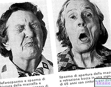
Huntington's disease is a genetic disease that, in its early stages, is characterized by loss of motor coordination and the presence of involuntary, exaggerated, jerking (chorea) movements or motor spasms involving the muscles of the limbs and face. The loss of motor control is progressive; at the same time, cognitive functions slowly decline until dementia.
The initial damage of Huntington's disease appears to be related to the degeneration of the nerve pathways that project from the basal ganglia to the thalamus.
Learn more: Huntington's disease (or Huntington's disease) - What it is, Causes, Symptoms and Diagnosis within the cerebral hemispheres. Also called basal nuclei or cerebral nuclei, the basal ganglia are found in each hemisphere below the lateral ventricles. The basal ganglia are surrounded by white matter and between or around them are fibers projection and commissures.
How are they organized?
From a functional point of view, the basal ganglia consist of the main components:
- Striatum (in turn, dorsal striatum - caudate and putamen nucleus - and ventral striatum - nucleus accumbens and olfactory tubercle);
- Globus pallidus;
- Substantia nigra (consisting in turn of a pars compacta and a pars reticulate);
- Subthalamic nucleus.
As for the striatum, it, in turn, is made up of the caudate nucleus, putamen and ventral striatum, which includes the nucleus accumbens and olfactory tubercle.
The striatum is important because it receives the main afferents of the nuclei of the base, from the cerebral cortex, from the thalamus and from the brain stem. Its neurons project to the globus pallidus and the substantia nigra.

From these two nuclei, whose neurons have morphologically similar somes or bodies, the main projections of the basal nuclei originate.
Note: Numerous other terms are used to indicate specific anatomical and functional subdivisions of the brain nuclei.
Basal ganglia: motor control
The basal ganglia are involved in subconscious control of muscle tone and coordination of learned movements. In practice, the basal ganglia ensure rhythm and correct voluntary movement patterns once this has begun.
The basal ganglia receive signals from the cortex and send them back to the cortex via intermediate synaptic stations in the thalamus. One of the functions of this neuronal circuit is to allow the cortex to optimize the initiation of intentional movements by inhibiting unwanted ones.
different. The striosome compartment receives afferents mainly from the limbic cortex and projects mainly to the pars compacta of the substantia nigra. To better understand the functioning of the striatum, it is appropriate to mention how the circuitry or the communication between the different brain areas works.
All areas of the cerebral cortex send glutamatergic excitatory projections to specific areas of the striatum. The striatum also receives excitatory signals from the intralaminar nuclei of the thalamus, dopaminergic projections from the midbrain and serotonergic projections from the raphe nuclei.
In particular, the striatum is made up of different cell types, but 90-95% of the cells that compose it are made up of GABAergic projection neurons. They represent the primary target of projections from the cerebral cortex and are also the sole source of efferent projections. They are normally silent neurons, except during movement or after the application of peripheral stimuli. The striatum is also made up of local inhibitory interneurons which, thanks to their developed axonal collaterals, reduce the activity of the efferent neurons of the striatum. Although these neurons are present in small quantities, most of the tonic activity of the striatum is due to them.
As for the circuitry, the striatum projects to the nuclei from which the efferent pathways originate through two pathways, a so-called direct path which is excitatory and an indirect path of the inhibitory type.
What are the functions of the striatum?
Through interactions with the cerebral cortex, the basal ganglia contribute to voluntary movement, but also to other forms of behavior such as skeleton-motor, oculomotor, cognitive and emotional functions.
For example, in some individuals with Huntington's disease, some lesions in the basal nuclei have been observed to produce negative emotional and cognitive effects.
damaged). In 1985, the scholar Vonsattel classified this disease from grade 0 (where no changes occur) to grade 4, in relation to the extent of atrophy of the striatum. It has also been shown that the degree of atrophy that occurs at the level of the striatum , also correlates with the degeneration of other non-striatal brain structures.
In the striatum the most affected neurons are the medium spiny neurons which represent the largest population present in the striatum and which use GABA, an inhibitory amino acid, as a neurotransmitter.
It has also been shown that Huntington's disease is characterized by the presence of intraneuronal inclusions and protein aggregates in dystrophic axons and in striatal and cortical neurons; this is also present in other polyglutamine disorders (i.e., triplet expansions, such as in the case of Huntington's disease). Furthermore, intranuclear inclusions have been shown to occur before brain weight loss, but also before body weight loss and before the onset of neurological symptoms.


