Generality
Syringomyelia is a neurological disease characterized by the formation of fluid-filled cysts in the spinal cord.

Figure: syringomyelia.
From the site: mdguidelines.com
In the long run, the presence of these cysts, called syringes, creates damage to the spinal cord; various ailments appear, such as pain in several parts of the body, a sense of stiffness and weakness, muscle atrophy, loss of reflexes, spasms in the legs, etc.
Syringomyelia can be due to different causes; in most cases, it is associated with a malformation of the cerebellum known as a Chiari malformation.
For a correct diagnosis, you need: a thorough physical examination of the patient, an analysis of his medical history and some diagnostic imaging tests.
The only way to empty the syringes and resolve (at least in part) the symptoms is to resort to surgery.
What is syringomyelia?
Syringomyelia is a rare pathological condition in which one or more fluid-filled cysts form within the spinal canal.
If cysts (also called syringes or fistulas) expand, they can severely damage the spinal cord and impair the transmission of nerve signals.
WHAT ARE CYSTS AND WHAT DO THEY CONTAIN?
Syringes are ducts or cavities (in Greek, "syrinx"means" conduit ") containing cerebrospinal fluid (or liquor).

Figure: the three meninges that surround the central nervous system.
The meninges are protective membranes: the most external is the dura mater; the central is the arachnoid, and finally, the innermost is the pia mater.
The subarachnoid space, between the arachnoid and the pia mater, contains the cerebrospinal fluid.
CSF is a colorless fluid that surrounds the central nervous system (or CNS), protects it from any trauma, provides it with nourishment and regulates its internal pressure (intracranial pressure).
The cephalorachidian fluid is produced inside the cerebral ventricles, precisely at the level of the chorioid plexuses; from here, it flows into the subarachnoid space, that is the area between the menynx pia mater and the arachnoid menynx.
EPIDEMIOLOGY
According to some statistical studies, syringomyelia would have a prevalence of about 8 cases per 100,000 people. Usually, its characteristic symptoms appear between the ages of 25 and 40 and its evolution is generally slow.
Many patients with syringomyelia also have a "structural alteration of the cerebellum, known as a Chiari malformation (or Arnold-Chiari or Arnold-Chiari syndrome).
Causes
Syringomyelia can be a congenital condition, that is present from birth, or acquired, that is, arisen in the course of life following a particular event or in association with a certain morbid state.
CONGENITAL SYRINGOMYELIA
Congenital forms of syringomyelia are due to Arnold-Chiari syndrome.
Arnold-Chiari syndrome is a cerebellar malformation (i.e. of the cerebellum) characterized by a downward displacement, precisely in the direction of the foramen magnum and the spinal canal, of the basal portion of the cerebellum.

In other words, it is a "cerebellar hernia, in which part of the cerebellum protrudes from the foramen magnum invading the canal that contains the spinal cord.
ACQUIRED SYRINGOMYELIA
The main causes that can lead to the onset of an acquired syringomyelia are:
- Trauma to the spinal cord. Among the various traumas that can lead to syringomyelia there is a fracture of the vertebrae.
- A meningitis. Meningitis are inflammations of the membranes surrounding the central nervous system (N.B: the CNS is composed of the brain and spinal cord). At the origin of a meningitis there can be a "viral or bacterial infection.
Arachnoiditis, ie inflammation of the arachnoid, is the form of meningitis that most frequently can cause syringomyelia. - A tumor in the spinal cord. According to experts, the presence of a tumor mass inside the spinal canal would prevent the normal circulation of the CSF, thus giving rise to a localized collection, then to a syringe.
- Stiff spine syndrome. It is a particular neurological disease, characterized by adhesions between the spinal cord and the vertebral column; adhesions that prevent the medulla from flowing normally.
The typical symptoms and signs of stiff spine syndrome are: leg pain, back pain, scoliosis, urinary incontinence or retention, muscle weakness and numbness etc. - A "bleeding inside" the spinal cord (or hematomyelia). Hematomyelia can be spontaneous or traumatic and usually affects the arterial vessels of the gray matter. Usually, the traumas that can result in hematomyelia are blows to the vertebrae.
Readers are reminded that some forms of syringomyelia arise without a definite cause or decipherable reason. In these situations, we speak of idiopathic acquired syringomyelia.
SUSPENDED POINTS
Doctors and researchers are still trying to solve some questions related to the formation of syringes, and how they damage the spinal cord.
According to some theories, it seems that the syringes originate from an "obstruction to the flow of CSF located in the subarachnoid space and that the damage to the spinal cord is the result of the pressure exerted by the cysts on the spinal cord itself.
Symptoms and Complications
For further information: Syringomyelia Symptoms
The classic signs and symptoms of syringomyelia are:
- Muscle weakness and atrophy
- Loss of reflexes
- Loss of sensitivity to pain and ambient temperature (N.B: in other words, the skin cannot feel the sensation of intense heat or freezing cold)
- Stiffness in the back, shoulders, arms and legs
- Pain in the neck, arms, hands and back
- Bowel and bladder problems. The patient loses control of the anal and bladder sphincters.
- Extreme muscle weakness and leg spasms
- Pain and numbness in the face
These manifestations, which at onset are generally not very marked, are the effect of the damage that the syringes produce to the spinal cord.
CONGENITAL SYRINGOMYELIA
Although present from birth, congenital syringes tend to cause the first symptoms in adulthood. The reason for this is explained by the fact that the cysts take a long time to produce significant damage to the spinal cord.
In addition to being affected by Chiari malformation, patients with congenital syringomyelia can also suffer from hydrocephalus and arachnoiditis.
What is hydrocephalus?
The term hydrocephalus indicates a serious disease due to an abnormal increase of the cerebrospinal fluid contained in the subarachnoid space and in the cerebral ventricles.
This disproportionate increase in CSF occurs when intracranial pressure has previously increased (intracranial hypertension). The cause of hydrocephalus can be: a brain tumor, a cerebral hemorrhage, meningitis, encephalitis, CNS malformations, etc.
The main signs of hydrocephalus are: increased head circumference, neck pain, epilepsy and convulsions.
ACQUIRED SYRINGOMYELIA
From the event that triggers the formation of syringes to the appearance of the first symptoms, months or years can pass. Therefore, similar to what happens in congenital syringomyelia, damage to the spinal cord progresses very slowly.
Typically, when trauma (post-traumatic syringomyelia) is the "origin c", the signs of the disease appear on one side of the body only.
WHEN TO SEE THE DOCTOR?
In the presence of one or more of the aforementioned symptoms, it is advisable to contact your doctor immediately to schedule an in-depth visit to identify the causes of the disorders.
Expert judgment is crucial, as syringomyelia is usually the result of other pre-existing diseases (which require appropriate treatment).
Those who have been the victim of severe back trauma may not initially experience any ailments related to syringomyelia. However, this could still develop after a certain period and become symptomatic even after a few months or years. Therefore, if any suspicious symptoms ever appear, it is advisable to contact your doctor and report the traumatic event suffered.
COMPLICATIONS
Syringomyelia is a potentially degenerative disease, as the syringes can widen, increasingly damage the spinal cord and even more importantly alter the patient's nervous functions.
Some classic expressions of these complications are:
- Scoliosis. It is an "abnormal curvature of the spine.
- Widespread chronic pain. The greater the damage to the spinal cord, the more intense and persistent the painful sensation in the neck, arms, hands and back becomes.
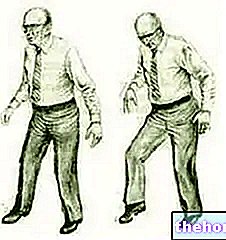
- Severe motor difficulties. If the atrophy and muscle weakness worsen, the sufferer may also have serious problems with walking.
Diagnosis
To diagnose syringomyelia, the first step is a physical examination and medical history analysis.
Therefore, once these two preliminary checks have been carried out, diagnostic imaging tests, such as nuclear magnetic resonance (NMR), CT scan, become of fundamental importance; for some patients, a lumbar puncture is also required.
OBJECTIVE EXAMINATION
During the physical examination, the doctor evaluates the symptomatic situation, asking the patient to describe in detail the complaints felt.
CLINICAL HISTORY EXAMINATION
When a doctor analyzes the medical history of a patient, he searches for possible triggers and possible situations predisposing the disease. For example, they are being investigated:
- Pathologies suffered by the patient in the past
- The pathologies in place at the time of the examination
- Recurrent pathologies within the family to which the patient belongs (a family member with similar disorders, etc.)
- Situations that have seen the patient suffer from spinal cord trauma.

Figure: Syringomyelia on nuclear magnetic resonance. The syringe is circled in red.
NUCLEAR MAGNETIC RESONANCE (NMR)
There are two different types of nuclear magnetic resonance (NMR):
- Classical magnetic resonance. By creating magnetic fields, MRI provides a "detailed image of the spinal cord, without exposing the patient to harmful ionizing radiation.
- Contrast MRI. The instrument used is the same as that used for classical MRI. The only difference is that the patient is injected with a contrast fluid, which serves to reveal the presence of any spinal cord tumors or other similar abnormalities.
The contrast liquid represents the only contraindication of the examination: this could in fact have toxic effects or trigger an allergy.
CT (COMPUTERIZED AXIAL TOMOGRAPHY)
Computed Axial Tomography (CT) provides clear images of internal organs, including the spinal cord. During its execution, the subject is exposed to a minimal amount of harmful ionizing radiation; therefore the test is to be considered, albeit minimally, invasive.
LUMBAR PUNCTURE
Lumbar puncture consists of taking a sample of cerebrospinal fluid and analyzing it in the laboratory. A needle inserted between the L3-L4 or L4-L5 lumbar vertebrae is used to withdraw the CSF.
Lumbar puncture is very useful for investigating the causes of syringomyelia: in fact, it can detect any infections in the meninges.
Treatment
When the symptoms of syringomyelia interfere with normal daily life, surgery is the only remedy.
Conversely, when syringomyelia does not cause any disturbance, simple monitoring of the situation is provided (principle of surveillance).
PRINCIPLE OF SURVEILLANCE
The principle of surveillance consists in subjecting the patient to periodic magnetic resonances and neurological control examinations. This approach is indicated for subjects with asymptomatic syringes (ie that do not cause any obvious symptoms).
Although this is a very remote hypothesis, it is possible that the cysts will disappear spontaneously.
SURGERY
The surgery to be adopted is chosen on the basis of the conditions that caused (or accompany) the syringomyelia. Therefore:
- In case of Chiari malformation, the operating surgeon can resort to the following operations: decompression of the posterior fossa, decompression of the spinal cord (decompressive laminectomy) and decompressive incision of the dura mater. All three procedures aim to reduce compression to damage the cerebellum and spinal cord by trying to increase the space available to them. More space available means an improvement in the flow of liquor, therefore a possible emptying of the syringes.
With the decompression of the posterior fossa, part of the posterior portion of the occipital bone is removed.
With decompressive laminectomy, the vertebral portion that delimits the hole through which the spinal cord passes is eliminated.
Finally, with the decompression incision of the dura mater, the outermost menynx is incised. - In case of spinal cord cancer, the surgeon eliminates the tumor mass, in such a way as to re-establish the flow of liquor. After removal, the syringe is automatically emptied, making the symptoms that characterized it before the intervention regress.
- In case of rigid spine syndrome, the surgeon can resort to various methods. The main one consists in "dissolving" the adhesions that block the column to the spinal cord.
Possible complications of syringoperitoneal shunt:
- Spinal cord injury
- Infections
- Fluid circulation blocks
- Bleeding within the spinal cord (hematomyelia)
Alongside these specific surgical techniques, there is also a procedure aimed at temporarily reducing symptoms and painful sensation (palliative therapy), known as a surgical shunt.
The surgical shunt is a drainage system, consisting of a flexible tube that allows the syringes to be emptied. In general, the excess fluid is drained into the abdomen (syringoperitoneal shunt), in a similar way to what is done in the case of hydrocephalus (ventriculoperitoneal shunt).
Since it is a rather delicate and not free from complications method, before putting it into practice the doctor carefully evaluates the situation and discusses the possible risks with the patient.
Who performs the surgeries?
The operating surgeon is a doctor who specializes in neurosurgery, which is the branch of surgery that specifically deals with problems and diseases of the central and peripheral nervous system.
CARE IN CASE OF RELAPSE
Even when properly treated, syringomyelia can recur after some time (relapse).
In order to promptly detect a "possible recurrence, it is advisable to periodically undergo appropriate medical checks and a nuclear magnetic resonance of the back.
The reformation of one or more syringes requires a second surgery, often to drain the cyst, but the benefits of which may only be temporary.
SOME ADVICES
Syringomyelia could severely affect the quality of life of the affected person and any of their daily activities. Therefore, it is a good idea:
- Avoid lifting weights, straining and "loading" your back, as they are three actions that can worsen symptoms, especially pain.
- Contact a good physiotherapist. Thanks to an "appropriate physiotherapy, it is possible to improve mobility, strengthen the muscles and withstand efforts better.
- Treat chronic pain with appropriate medications. If the pain is chronic and excruciating, the patient should ask their doctor to plan an "adequate pain relief therapy."
- Seek comfort from loved ones (friends or relatives) and join a support group for people with syringomyelia. There are two different ways to improve the mood, often depressed due to the limitations imposed by the disease.
Prognosis
The prognosis depends on the severity of the causes that triggered syringomyelia and on the effectiveness of the surgery.
- It depends on the severity of the triggers because, the more serious the condition that triggers syringomyelia, the more likely it is that the consequences of the disease are permanent.
- It depends on the effectiveness of the treatment since the medical intervention does not always restore the normal flow of liquor, just as it does not always allow the definitive elimination of the cysts associated with syringomyelia.


